 Movie
Movie Controller


+ Open data

- Basic information
| Entry | Database: PDB / ID: 8rrh | ||||||||||||||||||
|---|---|---|---|---|---|---|---|---|---|---|---|---|---|---|---|---|---|---|---|
| Title | The human prohibitin complex | ||||||||||||||||||
 Components Components |
| ||||||||||||||||||
 Keywords Keywords | MEMBRANE PROTEIN / Chaperone / Lipid organization / Protease regulator | ||||||||||||||||||
| Function / homology |  Function and homology information Function and homology informationregulation of cardiolipin metabolic process / mitochondrial prohibitin complex / : / complement component C3a binding / proteinase activated receptor binding / host-mediated perturbation of viral RNA genome replication / Processing of SMDT1 / sphingolipid binding / negative regulation of nuclear receptor-mediated glucocorticoid signaling pathway / Cellular response to mitochondrial stress ...regulation of cardiolipin metabolic process / mitochondrial prohibitin complex / : / complement component C3a binding / proteinase activated receptor binding / host-mediated perturbation of viral RNA genome replication / Processing of SMDT1 / sphingolipid binding / negative regulation of nuclear receptor-mediated glucocorticoid signaling pathway / Cellular response to mitochondrial stress / RIG-I signaling pathway / positive regulation of complement activation / T-helper 17 type immune response / complement component C3b binding / negative regulation of androgen receptor signaling pathway / : / positive regulation of G protein-coupled receptor signaling pathway / cellular response to interleukin-6 / positive regulation of interleukin-17 production / sister chromatid cohesion / DNA biosynthetic process / positive regulation of immunoglobulin production / progesterone receptor signaling pathway / B cell activation / mitophagy / positive regulation of smooth muscle cell proliferation / protein import into nucleus / nuclear estrogen receptor binding / antiviral innate immune response / cell periphery / mitochondrion organization / negative regulation of protein catabolic process / negative regulation of ERK1 and ERK2 cascade / epigenetic regulation of gene expression / RAF activation / negative regulation of cell growth / positive regulation of non-canonical NF-kappaB signal transduction / osteoblast differentiation / histone deacetylase binding / nuclear matrix / positive regulation of neuron apoptotic process / Signaling by moderate kinase activity BRAF mutants / Paradoxical activation of RAF signaling by kinase inactive BRAF / Signaling downstream of RAS mutants / transcription corepressor activity / cell migration / positive regulation of phosphatidylinositol 3-kinase/protein kinase B signal transduction / early endosome / regulation of apoptotic process / positive regulation of ERK1 and ERK2 cascade / mitochondrial outer membrane / protein stabilization / mitochondrial inner membrane / negative regulation of cell population proliferation / protein heterodimerization activity / negative regulation of DNA-templated transcription / positive regulation of gene expression / regulation of transcription by RNA polymerase II / negative regulation of apoptotic process / symbiont entry into host cell / regulation of DNA-templated transcription / positive regulation of DNA-templated transcription / negative regulation of transcription by RNA polymerase II / enzyme binding / cell surface / signal transduction / protein homodimerization activity / protein-containing complex / mitochondrion / DNA-templated transcription / extracellular exosome / nucleoplasm / membrane / identical protein binding / nucleus / plasma membrane / cytoplasm Similarity search - Function | ||||||||||||||||||
| Biological species |  Homo sapiens (human) Homo sapiens (human) | ||||||||||||||||||
| Method | ELECTRON MICROSCOPY / subtomogram averaging / cryo EM / Resolution: 16.3 Å | ||||||||||||||||||
 Authors Authors | Lange, F. / Ratz, M. / Dohrke, J.N. / Wenzel, D. / Riedel, D. / Ilgen, P. / Jakobs, S. | ||||||||||||||||||
| Funding support |  Germany, European Union, 5items Germany, European Union, 5items
| ||||||||||||||||||
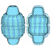
 Citation Citation | Journal: Nat Cell Biol / Year: 2025 Title: In situ architecture of the human prohibitin complex. Authors: Felix Lange / Michael Ratz / Jan-Niklas Dohrke / Maxence Le Vasseur / Dirk Wenzel / Peter Ilgen / Dietmar Riedel / Stefan Jakobs /   Abstract: Prohibitins are a highly conserved family of proteins that have been implicated in a variety of functions including mitochondrial stress signalling and housekeeping, cell cycle progression, ...Prohibitins are a highly conserved family of proteins that have been implicated in a variety of functions including mitochondrial stress signalling and housekeeping, cell cycle progression, apoptosis, lifespan regulation and many others. The human prohibitins prohibitin 1 and prohibitin 2 have been proposed to act as scaffolds within the mitochondrial inner membrane, but their molecular organization has remained elusive. Here we determined the molecular organization of the human prohibitin complex within the mitochondrial inner membrane using an integrative structural biology approach combining quantitative western blotting, cryo-electron tomography, subtomogram averaging and molecular modelling. The proposed bell-shaped structure consists of 11 alternating prohibitin 1 and prohibitin 2 molecules. This study reveals an average of about 43 prohibitin complexes per crista, covering 1-3% of the crista membrane area. These findings provide a structural basis for understanding the functional contributions of prohibitins to the integrity and spatial organization of the mitochondrial inner membrane. | ||||||||||||||||||
| History |
|
- Structure visualization
| Structure viewer | Molecule: MolmilJmol/JSmol |
|---|
- Downloads & links
-Download
| PDBx/mmCIF format | 8rrh.cif.gz | 535.9 KB | Display | PDBx/mmCIF format |
|---|---|---|---|---|
| PDB format | pdb8rrh.ent.gz | 441.7 KB | Display | PDB format |
| PDBx/mmJSON format | 8rrh.json.gz | Tree view | PDBx/mmJSON format | |
| Others |  Other downloads Other downloads |
-Validation report
| Arichive directory | https://data.pdbj.org/pub/pdb/validation_reports/rr/8rrhftp://data.pdbj.org/pub/pdb/validation_reports/rr/8rrh | HTTPS FTP |
|---|
-Related structure data
| Related structure data |  19459MC M: map data used to model this data C: citing same article ( |
|---|---|
| Similar structure data |
-Links
 PDBj
PDBj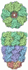
- Assembly
| Deposited unit | 
|
|---|---|
| 1 |
|
-Components
| #1: Protein | Mass: 29838.029 Da / Num. of mol.: 6 / Source method: isolated from a natural source / Source: (natural) Homo sapiens (human) / Cell line: U2OS / References: UniProt: P35232#2: Protein | Mass: 33341.355 Da / Num. of mol.: 5 / Source method: isolated from a natural source / Source: (natural) Homo sapiens (human) / References: UniProt: Q99623Has protein modification | N | |
|---|
-Experimental details
-Experiment
| Experiment | Method: ELECTRON MICROSCOPY |
|---|---|
| EM experiment | Aggregation state: CELL / 3D reconstruction method: subtomogram averaging |
- Sample preparation
| Component | Name: human prohibitin complex formed by PHB1 and PHB2 / Type: CELL Details: 11 molecules of PHB1 and PHB2 form the human prohibitin complex Entity ID: all / Source: NATURAL |
|---|---|
| Source (natural) | Organism: Homo sapiens (human) / Cellular location: mitochondrial inner membrane / Organelle: mitochondria |
| Buffer solution | pH: 7.4 / Details: DMEM high glucose medium (Gibco) |
| Specimen | Embedding applied: NO / Shadowing applied: NO / Staining applied: NO / Vitrification applied: YES |
| Specimen support | Grid material: GOLD / Grid mesh size: 200 divisions/in. / Grid type: Quantifoil R2/2 |
| Vitrification | Instrument: FEI VITROBOT MARK IV / Cryogen name: ETHANE / Humidity: 95 % / Chamber temperature: 277.15 K |
- Electron microscopy imaging
| Experimental equipment |  Model: Titan Krios / Image courtesy: FEI Company |
|---|---|
| Microscopy | Model: FEI TITAN KRIOS |
| Electron gun | Electron source:  FIELD EMISSION GUN / Accelerating voltage: 300 kV / Illumination mode: FLOOD BEAM FIELD EMISSION GUN / Accelerating voltage: 300 kV / Illumination mode: FLOOD BEAM |
| Electron lens | Mode: BRIGHT FIELD / Nominal magnification: 42000 X / Nominal defocus max: 4000 nm / Nominal defocus min: 2500 nm / Cs: 2.7 mm / C2 aperture diameter: 70 µm / Alignment procedure: COMA FREE |
| Specimen holder | Cryogen: NITROGEN / Specimen holder model: FEI TITAN KRIOS AUTOGRID HOLDER / Temperature (max): 93 K / Temperature (min): 80 K |
| Image recording | Average exposure time: 0.53 sec. / Electron dose: 120 e/Å2 / Avg electron dose per subtomogram: 120 e/Å2 / Film or detector model: GATAN K3 BIOQUANTUM (6k x 4k) / Num. of real images: 4 |
| EM imaging optics | Energyfilter name: GIF Bioquantum / Energyfilter slit width: 20 eV |
| Image scans | Width: 5760 / Height: 4092 |
- Processing
| EM software |
| ||||||||||||||||||||||||||||||||||||||||
|---|---|---|---|---|---|---|---|---|---|---|---|---|---|---|---|---|---|---|---|---|---|---|---|---|---|---|---|---|---|---|---|---|---|---|---|---|---|---|---|---|---|
| CTF correction | Type: PHASE FLIPPING AND AMPLITUDE CORRECTION | ||||||||||||||||||||||||||||||||||||||||
| Symmetry | Point symmetry: C11 (11 fold cyclic) | ||||||||||||||||||||||||||||||||||||||||
| 3D reconstruction | Resolution: 16.3 Å / Resolution method: FSC 0.143 CUT-OFF / Num. of particles: 817 / Algorithm: BACK PROJECTION / Num. of class averages: 1 / Symmetry type: POINT | ||||||||||||||||||||||||||||||||||||||||
| EM volume selection | Method: manual picking / Num. of tomograms: 37 / Num. of volumes extracted: 817 / Reference model: none | ||||||||||||||||||||||||||||||||||||||||
| Atomic model building | Protocol: FLEXIBLE FIT / Space: REAL | ||||||||||||||||||||||||||||||||||||||||
| Atomic model building | Source name: AlphaFold / Type: in silico model |
