 Movie
Movie Controller


+ Open data

- Basic information
| Entry | Database: PDB / ID: 8qp8 | ||||||
|---|---|---|---|---|---|---|---|
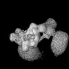
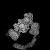
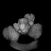
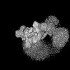
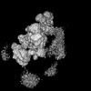
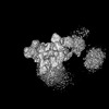
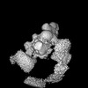
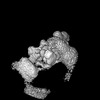
| Title | Cryo-EM Structure of Pre-B Complex (core part) | ||||||
 Components Components |
| ||||||
 Keywords Keywords | SPLICING / spliceosome | ||||||
| Function / homology |  Function and homology information Function and homology informationribonucleoprotein complex localization / U4atac snRNP / positive regulation of cytotoxic T cell differentiation / maturation of 5S rRNA / RNA localization / U4atac snRNA binding / R-loop processing / cis assembly of pre-catalytic spliceosome / negative regulation of mRNA splicing, via spliceosome / RNA splicing, via transesterification reactions ...ribonucleoprotein complex localization / U4atac snRNP / positive regulation of cytotoxic T cell differentiation / maturation of 5S rRNA / RNA localization / U4atac snRNA binding / R-loop processing / cis assembly of pre-catalytic spliceosome / negative regulation of mRNA splicing, via spliceosome / RNA splicing, via transesterification reactions / U2-type catalytic step 1 spliceosome / U4 snRNA binding / snRNP binding / spliceosomal tri-snRNP complex / U2-type precatalytic spliceosome / U2-type spliceosomal complex / mRNA cis splicing, via spliceosome / U2-type prespliceosome assembly / U2-type catalytic step 2 spliceosome / U4 snRNP / U2 snRNP / U2-type prespliceosome / K63-linked polyubiquitin modification-dependent protein binding / precatalytic spliceosome / spliceosomal complex assembly / mRNA Splicing - Minor Pathway / mRNA 3'-splice site recognition / MLL1 complex / spliceosomal tri-snRNP complex assembly / protein deubiquitination / U5 snRNA binding / U5 snRNP / U2 snRNA binding / U6 snRNA binding / pre-mRNA intronic binding / spliceosomal snRNP assembly / ribonucleoprotein complex binding / Cajal body / U1 snRNA binding / U4/U6 x U5 tri-snRNP complex / catalytic step 2 spliceosome / mRNA Splicing - Major Pathway / RNA splicing / response to cocaine / spliceosomal complex / mRNA splicing, via spliceosome / cellular response to xenobiotic stimulus / mRNA processing / cellular response to tumor necrosis factor / cellular response to lipopolysaccharide / protein-macromolecule adaptor activity / nucleic acid binding / ubiquitinyl hydrolase 1 / hydrolase activity / RNA helicase activity / nuclear speck / ciliary basal body / RNA helicase / cell division / mRNA binding / intracellular membrane-bounded organelle / GTPase activity / centrosome / chromatin / GTP binding / nucleolus / Golgi apparatus / positive regulation of transcription by RNA polymerase II / ATP hydrolysis activity / RNA binding / extracellular exosome / zinc ion binding / nucleoplasm / ATP binding / identical protein binding / nucleus / membrane / cytosol / cytoplasm Similarity search - Function | ||||||
| Biological species |  Homo sapiens (human) Homo sapiens (human) | ||||||
| Method | ELECTRON MICROSCOPY / single particle reconstruction / cryo EM / Resolution: 3.5 Å | ||||||
 Authors Authors | Zhang, Z. / Kumar, V. / Dybkov, O. / Will, C.L. / Zhong, J. / Ludwig, S. / Urlaub, H. / Kastner, B. / Stark, H. / Luehrmann, R. | ||||||
| Funding support |  Germany, 1items Germany, 1items
| ||||||
 Citation Citation | Journal: Nature / Year: 2024 Title: Structural insights into the cross-exon to cross-intron spliceosome switch. Authors: Zhenwei Zhang / Vinay Kumar / Olexandr Dybkov / Cindy L Will / Jiayun Zhong / Sebastian E J Ludwig / Henning Urlaub / Berthold Kastner / Holger Stark / Reinhard Lührmann /  Abstract: Early spliceosome assembly can occur through an intron-defined pathway, whereby U1 and U2 small nuclear ribonucleoprotein particles (snRNPs) assemble across the intron. Alternatively, it can occur ...Early spliceosome assembly can occur through an intron-defined pathway, whereby U1 and U2 small nuclear ribonucleoprotein particles (snRNPs) assemble across the intron. Alternatively, it can occur through an exon-defined pathway, whereby U2 binds the branch site located upstream of the defined exon and U1 snRNP interacts with the 5' splice site located directly downstream of it. The U4/U6.U5 tri-snRNP subsequently binds to produce a cross-intron (CI) or cross-exon (CE) pre-B complex, which is then converted to the spliceosomal B complex. Exon definition promotes the splicing of upstream introns and plays a key part in alternative splicing regulation. However, the three-dimensional structure of exon-defined spliceosomal complexes and the molecular mechanism of the conversion from a CE-organized to a CI-organized spliceosome, a pre-requisite for splicing catalysis, remain poorly understood. Here cryo-electron microscopy analyses of human CE pre-B complex and B-like complexes reveal extensive structural similarities with their CI counterparts. The results indicate that the CE and CI spliceosome assembly pathways converge already at the pre-B stage. Add-back experiments using purified CE pre-B complexes, coupled with cryo-electron microscopy, elucidate the order of the extensive remodelling events that accompany the formation of B complexes and B-like complexes. The molecular triggers and roles of B-specific proteins in these rearrangements are also identified. We show that CE pre-B complexes can productively bind in trans to a U1 snRNP-bound 5' splice site. Together, our studies provide new mechanistic insights into the CE to CI switch during spliceosome assembly and its effect on pre-mRNA splice site pairing at this stage. | ||||||
| History |
|
- Structure visualization
| Structure viewer | Molecule: MolmilJmol/JSmol |
|---|
- Downloads & links
-Download
| PDBx/mmCIF format | 8qp8.cif.gz | 891.6 KB | Display | PDBx/mmCIF format |
|---|---|---|---|---|
| PDB format | pdb8qp8.ent.gz | 644 KB | Display | PDB format |
| PDBx/mmJSON format | 8qp8.json.gz | Tree view | PDBx/mmJSON format | |
| Others |  Other downloads Other downloads |
-Validation report
| Summary document | 8qp8_validation.pdf.gz | 1.4 MB | Display | wwPDB validaton report |
|---|---|---|---|---|
| Full document | 8qp8_full_validation.pdf.gz | 1.5 MB | Display | |
| Data in XML | 8qp8_validation.xml.gz | 121 KB | Display | |
| Data in CIF | 8qp8_validation.cif.gz | 187.1 KB | Display | |
| Arichive directory | https://data.pdbj.org/pub/pdb/validation_reports/qp/8qp8ftp://data.pdbj.org/pub/pdb/validation_reports/qp/8qp8 | HTTPS FTP |
-Related structure data
| Related structure data |  18544MC  8qozC  8qp9C  8qpaC  8qpbC 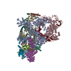 8qpeC  8qpkC 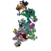 8qxdC 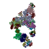 8qzsC 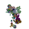 8r08C  8r09C  8r0aC  8r0bC  8rm5C M: map data used to model this data C: citing same article ( |
|---|---|
| Similar structure data |

-Links
 PDBj
PDBj


- Assembly
| Deposited unit | 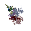
|
|---|---|
| 1 |
|
-Components
-Protein , 10 types, 10 molecules 7GSCUARNDX
| #1: Protein | Mass: 88991.094 Da / Num. of mol.: 1 / Source method: isolated from a natural source / Source: (natural) Homo sapiens (human) / References: UniProt: Q15459 |
|---|---|
| #2: Protein | Mass: 95785.148 Da / Num. of mol.: 1 / Source method: isolated from a natural source / Source: (natural) Homo sapiens (human) / References: UniProt: Q9BUQ8 |
| #6: Protein | Mass: 90414.117 Da / Num. of mol.: 1 / Source method: isolated from a natural source / Source: (natural) Homo sapiens (human) / References: UniProt: O43290 |
| #7: Protein | Mass: 109560.625 Da / Num. of mol.: 1 / Source method: isolated from a natural source / Source: (natural) Homo sapiens (human) / References: UniProt: Q15029 |
| #8: Protein | Mass: 65481.426 Da / Num. of mol.: 1 / Source method: isolated from a natural source / Source: (natural) Homo sapiens (human) / References: UniProt: Q53GS9, ubiquitinyl hydrolase 1 |
| #9: Protein | Mass: 273974.250 Da / Num. of mol.: 1 / Source method: isolated from a natural source / Source: (natural) Homo sapiens (human) / References: UniProt: Q6P2Q9 |
| #10: Protein | Mass: 50477.922 Da / Num. of mol.: 1 / Source method: isolated from a natural source / Source: (natural) Homo sapiens (human) / References: UniProt: Q9BTD8 |
| #11: Protein | Mass: 107092.242 Da / Num. of mol.: 1 / Source method: isolated from a natural source / Source: (natural) Homo sapiens (human) / References: UniProt: O94906 |
| #14: Protein | Mass: 16807.346 Da / Num. of mol.: 1 / Source method: isolated from a natural source / Source: (natural) Homo sapiens (human) / References: UniProt: P83876 |
| #15: Protein | Mass: 18915.414 Da / Num. of mol.: 1 / Source method: isolated from a natural source / Source: (natural) Homo sapiens (human) / References: UniProt: Q8WVK2 |
-RNA chain , 3 types, 3 molecules 654
| #3: RNA chain | Mass: 34098.270 Da / Num. of mol.: 1 / Source method: isolated from a natural source / Source: (natural) Homo sapiens (human) / References: GenBank: NR_004394.1 |
|---|---|
| #4: RNA chain | Mass: 37254.855 Da / Num. of mol.: 1 / Source method: isolated from a natural source / Source: (natural) Homo sapiens (human) / References: GenBank: 20330981 |
| #5: RNA chain | Mass: 46181.289 Da / Num. of mol.: 1 / Source method: isolated from a natural source / Source: (natural) Homo sapiens (human) / References: GenBank: 340142 |
-U4/U6 small nuclear ribonucleoprotein ... , 2 types, 2 molecules LJ
| #12: Protein | Mass: 55528.969 Da / Num. of mol.: 1 / Source method: isolated from a natural source / Source: (natural) Homo sapiens (human) / References: UniProt: Q8WWY3 |
|---|---|
| #13: Protein | Mass: 77669.188 Da / Num. of mol.: 1 / Source method: isolated from a natural source / Source: (natural) Homo sapiens (human) / References: UniProt: O43395 |
-Non-polymers , 1 types, 1 molecules 
| #16: Chemical | ChemComp-IHP /  Mass: 660.035 Da / Num. of mol.: 1 / Source method: obtained synthetically / Formula: C6H18O24P6 / Feature type: SUBJECT OF INVESTIGATION Mass: 660.035 Da / Num. of mol.: 1 / Source method: obtained synthetically / Formula: C6H18O24P6 / Feature type: SUBJECT OF INVESTIGATION |
|---|
-Details
| Has ligand of interest | Y |
|---|
-Experimental details
-Experiment
| Experiment | Method: ELECTRON MICROSCOPY |
|---|---|
| EM experiment | Aggregation state: PARTICLE / 3D reconstruction method: single particle reconstruction |
- Sample preparation
| Component | Name: human spliceosomal cross-exon pre-B complex / Type: COMPLEX / Entity ID: #1-#8, #11-#12, #14, #9-#10, #13, #15 / Source: NATURAL |
|---|---|
| Molecular weight | Experimental value: NO |
| Source (natural) | Organism: Homo sapiens (human) |
| Buffer solution | pH: 7.9 |
| Specimen | Embedding applied: NO / Shadowing applied: NO / Staining applied: NO / Vitrification applied: YES |
| Vitrification | Cryogen name: ETHANE |
- Electron microscopy imaging
| Experimental equipment |  Model: Titan Krios / Image courtesy: FEI Company |
|---|---|
| Microscopy | Model: FEI TITAN KRIOS |
| Electron gun | Electron source:  FIELD EMISSION GUN / Accelerating voltage: 300 kV / Illumination mode: FLOOD BEAM FIELD EMISSION GUN / Accelerating voltage: 300 kV / Illumination mode: FLOOD BEAM |
| Electron lens | Mode: BRIGHT FIELD / Nominal defocus max: 5000 nm / Nominal defocus min: 1500 nm |
| Image recording | Electron dose: 45 e/Å2 / Film or detector model: FEI FALCON III (4k x 4k) |
- Processing
| CTF correction | Type: PHASE FLIPPING AND AMPLITUDE CORRECTION |
|---|---|
| 3D reconstruction | Resolution: 3.5 Å / Resolution method: FSC 0.143 CUT-OFF / Num. of particles: 279781 / Symmetry type: POINT |
