 Movie
Movie Controller

[English] 日本語
 Yorodumi
- PDB-8q7n: cryo-EM structure of the human spliceosomal B complex protomer (t... -
+ Open data

- Basic information
| Entry | Database: PDB / ID: 8q7n | ||||||
|---|---|---|---|---|---|---|---|
| Title | cryo-EM structure of the human spliceosomal B complex protomer (tri-snRNP core region) | ||||||
 Components Components |
| ||||||
 Keywords Keywords | SPLICING / spliceosome / pre-catalytic spliceosome / spliceosomal B complex | ||||||
| Function / homology |  Function and homology information Function and homology informationmicrofibril / spliceosomal snRNP complex / ribonucleoprotein complex localization / U4atac snRNP / positive regulation of cytotoxic T cell differentiation / maturation of 5S rRNA / RNA localization / U4atac snRNA binding / box C/D sno(s)RNA binding / dense fibrillar component ...microfibril / spliceosomal snRNP complex / ribonucleoprotein complex localization / U4atac snRNP / positive regulation of cytotoxic T cell differentiation / maturation of 5S rRNA / RNA localization / U4atac snRNA binding / box C/D sno(s)RNA binding / dense fibrillar component / box C/D methylation guide snoRNP complex / U4/U6 snRNP / U2-type catalytic step 1 spliceosome / RNA splicing, via transesterification reactions / proline-rich region binding / spliceosomal tri-snRNP complex / snRNP binding / U2-type precatalytic spliceosome / mRNA cis splicing, via spliceosome / RNA polymerase binding / U2-type prespliceosome assembly / U2-type catalytic step 2 spliceosome / U2-type spliceosomal complex / U4 snRNA binding / transcription elongation factor activity / box C/D snoRNP assembly / U2 snRNP / U4 snRNP / U2-type prespliceosome / : / U3 snoRNA binding / K63-linked polyubiquitin modification-dependent protein binding / precatalytic spliceosome / rRNA modification in the nucleus and cytosol / ubiquitin-like protein conjugating enzyme binding / mRNA 3'-splice site recognition / mRNA Splicing - Minor Pathway / spliceosomal complex assembly / spliceosomal tri-snRNP complex assembly / negative regulation of transcription elongation by RNA polymerase II / MLL1 complex / U5 snRNP / U5 snRNA binding / pre-mRNA intronic binding / spliceosomal snRNP assembly / U2 snRNA binding / U6 snRNA binding / single fertilization / ribonucleoprotein complex binding / Cajal body / U1 snRNA binding / RNA processing / U4/U6 x U5 tri-snRNP complex / Major pathway of rRNA processing in the nucleolus and cytosol / catalytic step 2 spliceosome / mRNA Splicing - Major Pathway / RNA splicing / nuclear receptor binding / spliceosomal complex / transcription coregulator activity / response to cocaine / maturation of SSU-rRNA / positive regulation of transcription elongation by RNA polymerase II / small-subunit processome / mRNA splicing, via spliceosome / cellular response to xenobiotic stimulus / cellular response to tumor necrosis factor / mRNA processing / protein tag activity / transcription corepressor activity / ATPase binding / microtubule cytoskeleton / cellular response to lipopolysaccharide / ribosomal small subunit biogenesis / RNA polymerase II-specific DNA-binding transcription factor binding / protein-macromolecule adaptor activity / transcription coactivator activity / nuclear speck / ciliary basal body / cell division / GTPase activity / centrosome / GTP binding / chromatin / nucleolus / negative regulation of transcription by RNA polymerase II / Golgi apparatus / positive regulation of transcription by RNA polymerase II / protein-containing complex / DNA binding / RNA binding / zinc ion binding / nucleoplasm / membrane / identical protein binding / nucleus / cytoplasm / cytosol Similarity search - Function | ||||||
| Biological species |  Homo sapiens (human) Homo sapiens (human) | ||||||
| Method | ELECTRON MICROSCOPY / single particle reconstruction / cryo EM / Resolution: 3.1 Å | ||||||
 Authors Authors | Zhang, Z. / Kumar, V. / Dybkov, O. / Will, C.L. / Urlaub, H. / Stark, H. / Luehrmann, R. | ||||||
| Funding support |  Germany, 1items Germany, 1items
| ||||||
 Citation Citation | Journal: EMBO J / Year: 2024 Title: Cryo-EM analyses of dimerized spliceosomes provide new insights into the functions of B complex proteins. Authors: Zhenwei Zhang / Vinay Kumar / Olexandr Dybkov / Cindy L Will / Henning Urlaub / Holger Stark / Reinhard Lührmann /  Abstract: The B complex is a key intermediate stage of spliceosome assembly. To improve the structural resolution of monomeric, human spliceosomal B (hB) complexes and thereby generate a more comprehensive hB ...The B complex is a key intermediate stage of spliceosome assembly. To improve the structural resolution of monomeric, human spliceosomal B (hB) complexes and thereby generate a more comprehensive hB molecular model, we determined the cryo-EM structure of B complex dimers formed in the presence of ATP S. The enhanced resolution of these complexes allows a finer molecular dissection of how the 5' splice site (5'ss) is recognized in hB, and new insights into molecular interactions of FBP21, SNU23 and PRP38 with the U6/5'ss helix and with each other. It also reveals that SMU1 and RED are present as a heterotetrameric complex and are located at the interface of the B dimer protomers. We further show that MFAP1 and UBL5 form a 5' exon binding channel in hB, and elucidate the molecular contacts stabilizing the 5' exon at this stage. Our studies thus yield more accurate models of protein and RNA components of hB complexes. They further allow the localization of additional proteins and protein domains (such as SF3B6, BUD31 and TCERG1) whose position was not previously known, thereby uncovering new functions for B-specific and other hB proteins during pre-mRNA splicing. | ||||||
| History |
|
- Structure visualization
| Structure viewer | Molecule: MolmilJmol/JSmol |
|---|
- Downloads & links
-Download
| PDBx/mmCIF format | 8q7n.cif.gz | 1.5 MB | Display | PDBx/mmCIF format |
|---|---|---|---|---|
| PDB format | pdb8q7n.ent.gz | 1.1 MB | Display | PDB format |
| PDBx/mmJSON format | 8q7n.json.gz | Tree view | PDBx/mmJSON format | |
| Others |  Other downloads Other downloads |
-Validation report
| Arichive directory | https://data.pdbj.org/pub/pdb/validation_reports/q7/8q7nftp://data.pdbj.org/pub/pdb/validation_reports/q7/8q7n | HTTPS FTP |
|---|
-Related structure data
| Related structure data |  18225MC 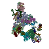 8qo9C M: map data used to model this data C: citing same article ( |
|---|---|
| Similar structure data |

-Links
 PDBj
PDBj


- Assembly
| Deposited unit | 
|
|---|---|
| 1 |
|
-Components
-RNA chain , 4 types, 4 molecules 56Z4
| #1: RNA chain | Mass: 37254.855 Da / Num. of mol.: 1 / Source method: isolated from a natural source / Source: (natural) Homo sapiens (human) / References: GenBank: 20330981 |
|---|---|
| #2: RNA chain | Mass: 34098.270 Da / Num. of mol.: 1 / Source method: isolated from a natural source / Source: (natural) Homo sapiens (human) / References: GenBank: NR_004394.1 |
| #11: RNA chain | Mass: 111300.453 Da / Num. of mol.: 1 / Source method: isolated from a natural source / Source: (natural) Homo sapiens (human) |
| #20: RNA chain | Mass: 46528.465 Da / Num. of mol.: 1 / Source method: isolated from a natural source / Source: (natural) Homo sapiens (human) |
-Protein , 14 types, 14 molecules 7CDIKMQXrsNAST
| #3: Protein | Mass: 88991.094 Da / Num. of mol.: 1 / Source method: isolated from a natural source / Source: (natural) Homo sapiens (human) / References: UniProt: Q15459 |
|---|---|
| #4: Protein | Mass: 109560.625 Da / Num. of mol.: 1 / Source method: isolated from a natural source / Source: (natural) Homo sapiens (human) / References: UniProt: Q15029 |
| #5: Protein | Mass: 16807.346 Da / Num. of mol.: 1 / Source method: isolated from a natural source / Source: (natural) Homo sapiens (human) / References: UniProt: P83876 |
| #6: Protein | Mass: 37563.863 Da / Num. of mol.: 1 / Source method: isolated from a natural source / Source: (natural) Homo sapiens (human) / References: UniProt: Q8NAV1 |
| #7: Protein | Mass: 52050.527 Da / Num. of mol.: 1 / Source method: isolated from a natural source / Source: (natural) Homo sapiens (human) / References: UniProt: P55081 |
| #8: Protein | Mass: 14191.524 Da / Num. of mol.: 1 / Source method: isolated from a natural source / Source: (natural) Homo sapiens (human) / References: UniProt: P55769 |
| #9: Protein | Mass: 17032.850 Da / Num. of mol.: 1 / Source method: isolated from a natural source / Source: (natural) Homo sapiens (human) / References: UniProt: P41223 |
| #10: Protein | Mass: 42575.801 Da / Num. of mol.: 1 / Source method: isolated from a natural source / Source: (natural) Homo sapiens (human) / References: UniProt: O75554 |
| #12: Protein | Mass: 23664.047 Da / Num. of mol.: 1 / Source method: isolated from a natural source / Source: (natural) Homo sapiens (human) / References: UniProt: Q96NC0 |
| #13: Protein | Mass: 8560.945 Da / Num. of mol.: 1 / Source method: isolated from a natural source / Source: (natural) Homo sapiens (human) / References: UniProt: Q9BZL1 |
| #16: Protein | Mass: 107092.242 Da / Num. of mol.: 1 / Source method: isolated from a natural source / Source: (natural) Homo sapiens (human) / References: UniProt: O94906 |
| #17: Protein | Mass: 273974.250 Da / Num. of mol.: 1 / Source method: isolated from a natural source / Source: (natural) Homo sapiens (human) / References: UniProt: Q6P2Q9 |
| #18: Protein | Mass: 90414.117 Da / Num. of mol.: 1 / Source method: isolated from a natural source / Source: (natural) Homo sapiens (human) / References: UniProt: O43290 |
| #19: Protein | Mass: 124083.000 Da / Num. of mol.: 1 / Source method: isolated from a natural source / Source: (natural) Homo sapiens (human) / References: UniProt: O14776 |
-U4/U6 small nuclear ribonucleoprotein ... , 3 types, 3 molecules LFJ
| #14: Protein | Mass: 55528.969 Da / Num. of mol.: 1 / Source method: isolated from a natural source / Source: (natural) Homo sapiens (human) / References: UniProt: Q8WWY3 |
|---|---|
| #15: Protein | Mass: 58536.105 Da / Num. of mol.: 1 / Source method: isolated from a natural source / Source: (natural) Homo sapiens (human) / References: UniProt: O43172 |
| #21: Protein | Mass: 77669.188 Da / Num. of mol.: 1 / Source method: isolated from a natural source / Source: (natural) Homo sapiens (human) / References: UniProt: O43395 |
-Details
| Has protein modification | Y |
|---|
-Experimental details
-Experiment
| Experiment | Method: ELECTRON MICROSCOPY |
|---|---|
| EM experiment | Aggregation state: PARTICLE / 3D reconstruction method: single particle reconstruction |
- Sample preparation
| Component | Name: human pre-catalytic spliceosome / Type: COMPLEX / Entity ID: #1-#11, #13-#20, #12, #21 / Source: NATURAL |
|---|---|
| Source (natural) | Organism: Homo sapiens (human) |
| Buffer solution | pH: 7.9 |
| Specimen | Embedding applied: NO / Shadowing applied: NO / Staining applied: NO / Vitrification applied: YES |
| Vitrification | Cryogen name: ETHANE |
- Electron microscopy imaging
| Experimental equipment |  Model: Titan Krios / Image courtesy: FEI Company |
|---|---|
| Microscopy | Model: FEI TITAN KRIOS |
| Electron gun | Electron source:  FIELD EMISSION GUN / Accelerating voltage: 300 kV / Illumination mode: FLOOD BEAM FIELD EMISSION GUN / Accelerating voltage: 300 kV / Illumination mode: FLOOD BEAM |
| Electron lens | Mode: BRIGHT FIELD / Nominal defocus max: 3000 nm / Nominal defocus min: 1500 nm |
| Image recording | Electron dose: 48 e/Å2 / Detector mode: INTEGRATING / Film or detector model: FEI FALCON III (4k x 4k) |
- Processing
| CTF correction | Type: PHASE FLIPPING AND AMPLITUDE CORRECTION |
|---|---|
| 3D reconstruction | Resolution: 3.1 Å / Resolution method: FSC 0.143 CUT-OFF / Num. of particles: 251564 / Symmetry type: POINT |
