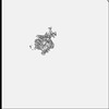
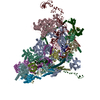
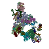
Journal: EMBO J / Year: 2024 Title: Cryo-EM analyses of dimerized spliceosomes provide new insights into the functions of B complex proteins. Authors: Zhenwei Zhang / Vinay Kumar / Olexandr Dybkov / Cindy L Will / Henning Urlaub / Holger Stark / Reinhard Lührmann / Abstract: The B complex is a key intermediate stage of spliceosome assembly. To improve the structural resolution of monomeric, human spliceosomal B (hB) complexes and thereby generate a more comprehensive hB ...The B complex is a key intermediate stage of spliceosome assembly. To improve the structural resolution of monomeric, human spliceosomal B (hB) complexes and thereby generate a more comprehensive hB molecular model, we determined the cryo-EM structure of B complex dimers formed in the presence of ATP S. The enhanced resolution of these complexes allows a finer molecular dissection of how the 5' splice site (5'ss) is recognized in hB, and new insights into molecular interactions of FBP21, SNU23 and PRP38 with the U6/5'ss helix and with each other. It also reveals that SMU1 and RED are present as a heterotetrameric complex and are located at the interface of the B dimer protomers. We further show that MFAP1 and UBL5 form a 5' exon binding channel in hB, and elucidate the molecular contacts stabilizing the 5' exon at this stage. Our studies thus yield more accurate models of protein and RNA components of hB complexes. They further allow the localization of additional proteins and protein domains (such as SF3B6, BUD31 and TCERG1) whose position was not previously known, thereby uncovering new functions for B-specific and other hB proteins during pre-mRNA splicing.
In the structure databanks used in Yorodumi, some data are registered as the other names, "COVID-19 virus" and "2019-nCoV". Here are the details of the virus and the list of structure data.
Jan 31, 2019. EMDB accession codes are about to change! (news from PDBe EMDB page)
EMDB accession codes are about to change! (news from PDBe EMDB page)
The allocation of 4 digits for EMDB accession codes will soon come to an end. Whilst these codes will remain in use, new EMDB accession codes will include an additional digit and will expand incrementally as the available range of codes is exhausted. The current 4-digit format prefixed with “EMD-” (i.e. EMD-XXXX) will advance to a 5-digit format (i.e. EMD-XXXXX), and so on. It is currently estimated that the 4-digit codes will be depleted around Spring 2019, at which point the 5-digit format will come into force.
The EM Navigator/Yorodumi systems omit the EMD- prefix.
Related info.:Q: What is EMD? / ID/Accession-code notation in Yorodumi/EM Navigator
Yorodumi is a browser for structure data from EMDB, PDB, SASBDB, etc.
This page is also the successor to EM Navigator detail page, and also detail information page/front-end page for Omokage search.
The word "yorodu" (or yorozu) is an old Japanese word meaning "ten thousand". "mi" (miru) is to see.
Related info.:EMDB / PDB / SASBDB / Comparison of 3 databanks / Yorodumi Search / Aug 31, 2016. New EM Navigator & Yorodumi / Yorodumi Papers / Jmol/JSmol / Function and homology information / Changes in new EM Navigator and Yorodumi
 Movie
Movie Controller


 Open data
 Basic information
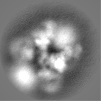
 Map data
 Sample
 Keywords
 Homo sapiens (human)
 Authors
 Germany, 1 items
  Citation
 Structure visualization
 Downloads & links
 EMDB map data format
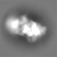
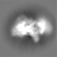
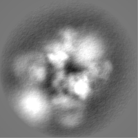
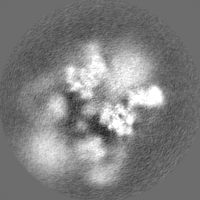
 emd_19063.png
 http://ftp.pdbj.org/pub/emdb/structures/EMD-19063
 Z (Sec.)
 Y (Row.)
 X (Col.)


 Sample components
 Processing
 Electron microscopy
 FIELD EMISSION GUN