 Movie
Movie Controller

[English] 日本語
 Yorodumi
- PDB-8ovx: Cryo-EM structure of yeast CENP-OPQU+ bound to the CENP-A N-terminus -
+ Open data

- Basic information
| Entry | Database: PDB / ID: 8ovx | |||||||||||||||||||||||||||||||||
|---|---|---|---|---|---|---|---|---|---|---|---|---|---|---|---|---|---|---|---|---|---|---|---|---|---|---|---|---|---|---|---|---|---|---|
| Title | Cryo-EM structure of yeast CENP-OPQU+ bound to the CENP-A N-terminus | |||||||||||||||||||||||||||||||||
 Components Components | (Inner kinetochore subunit ...) x 6 | |||||||||||||||||||||||||||||||||
 Keywords Keywords | CELL CYCLE / kinetochore / point centromere / CENP-A nucleosome / topological entrapment / centromeric DNA | |||||||||||||||||||||||||||||||||
| Function / homology |  Function and homology information Function and homology informationnegative regulation of meiotic DNA double-strand break formation involved in reciprocal meiotic recombination / COMA complex / Mis6-Sim4 complex / attachment of spindle microtubules to kinetochore / protein localization to chromosome, centromeric region / establishment of mitotic sister chromatid cohesion / protein localization to kinetochore / spindle pole body / mitotic spindle assembly checkpoint signaling / meiotic cell cycle ...negative regulation of meiotic DNA double-strand break formation involved in reciprocal meiotic recombination / COMA complex / Mis6-Sim4 complex / attachment of spindle microtubules to kinetochore / protein localization to chromosome, centromeric region / establishment of mitotic sister chromatid cohesion / protein localization to kinetochore / spindle pole body / mitotic spindle assembly checkpoint signaling / meiotic cell cycle / chromosome segregation / kinetochore / cell division / nucleus / cytoplasm Similarity search - Function | |||||||||||||||||||||||||||||||||
| Biological species |  | |||||||||||||||||||||||||||||||||
| Method | ELECTRON MICROSCOPY / single particle reconstruction / cryo EM / Resolution: 3.4 Å | |||||||||||||||||||||||||||||||||
 Authors Authors | Dendooven, T.D. / Zhang, Z. / Yang, J. / McLaughlin, S. / Schwabb, J. / Scheres, S. / Yatskevich, S. | |||||||||||||||||||||||||||||||||
| Funding support |  United Kingdom, United Kingdom,  Germany, 4items Germany, 4items
| |||||||||||||||||||||||||||||||||
 Citation Citation | Journal: Sci Adv / Year: 2023 Title: Cryo-EM structure of the complete inner kinetochore of the budding yeast point centromere. Authors: Tom Dendooven / Ziguo Zhang / Jing Yang / Stephen H McLaughlin / Johannes Schwab / Sjors H W Scheres / Stanislau Yatskevich / David Barford / Abstract: The point centromere of budding yeast specifies assembly of the large kinetochore complex to mediate chromatid segregation. Kinetochores comprise the centromere-associated inner kinetochore (CCAN) ...The point centromere of budding yeast specifies assembly of the large kinetochore complex to mediate chromatid segregation. Kinetochores comprise the centromere-associated inner kinetochore (CCAN) complex and the microtubule-binding outer kinetochore KNL1-MIS12-NDC80 (KMN) network. The budding yeast inner kinetochore also contains the DNA binding centromere-binding factor 1 (CBF1) and CBF3 complexes. We determined the cryo-electron microscopy structure of the yeast inner kinetochore assembled onto the centromere-specific centromere protein A nucleosomes (CENP-A). This revealed a central CENP-A with extensively unwrapped DNA ends. These free DNA duplexes bind two CCAN protomers, one of which entraps DNA topologically, positioned on the centromere DNA element I (CDEI) motif by CBF1. The two CCAN protomers are linked through CBF3 forming an arch-like configuration. With a structural mechanism for how CENP-A can also be linked to KMN involving only CENP-QU, we present a model for inner kinetochore assembly onto a point centromere and how it organizes the outer kinetochore for chromosome attachment to the mitotic spindle. | |||||||||||||||||||||||||||||||||
| History |
|
- Structure visualization
| Structure viewer | Molecule: MolmilJmol/JSmol |
|---|
- Downloads & links
-Download
| PDBx/mmCIF format | 8ovx.cif.gz | 184.5 KB | Display | PDBx/mmCIF format |
|---|---|---|---|---|
| PDB format | pdb8ovx.ent.gz | 131.3 KB | Display | PDB format |
| PDBx/mmJSON format | 8ovx.json.gz | Tree view | PDBx/mmJSON format | |
| Others |  Other downloads Other downloads |
-Validation report
| Arichive directory | https://data.pdbj.org/pub/pdb/validation_reports/ov/8ovxftp://data.pdbj.org/pub/pdb/validation_reports/ov/8ovx | HTTPS FTP |
|---|
-Related structure data
| Related structure data |  17225MC 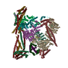 8ovwC 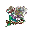 8ow0C 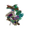 8ow1C M: map data used to model this data C: citing same article ( |
|---|---|
| Similar structure data |


-Links
 PDBj
PDBj- Assembly
| Deposited unit | 
|
|---|---|
| 1 |
|
-Components
-Inner kinetochore subunit ... , 6 types, 6 molecules OPQUYZ
| #1: Protein | Mass: 43028.879 Da / Num. of mol.: 1 Source method: isolated from a genetically manipulated source Source: (gene. exp.) Gene: MCM21, CTF5, YDR318W / Production host:  Trichoplusia ni (cabbage looper) / References: UniProt: Q06675 Trichoplusia ni (cabbage looper) / References: UniProt: Q06675 |
|---|---|
| #2: Protein | Mass: 42841.113 Da / Num. of mol.: 1 Source method: isolated from a genetically manipulated source Source: (gene. exp.) Gene: CTF19, MCM18, YPL018W, LPB13W / Production host: Trichoplusia ni (cabbage looper) / References: UniProt: Q02732 |
| #3: Protein | Mass: 47427.246 Da / Num. of mol.: 1 Source method: isolated from a genetically manipulated source Source: (gene. exp.) Gene: OKP1, YGR179C / Production host: Trichoplusia ni (cabbage looper) / References: UniProt: P53298 |
| #4: Protein | Mass: 37506.723 Da / Num. of mol.: 1 Source method: isolated from a genetically manipulated source Source: (gene. exp.) Gene: AME1, ARP100, YBR211C, YBR1458 / Production host: Trichoplusia ni (cabbage looper) / References: UniProt: P38313 |
| #5: Protein | Mass: 27006.451 Da / Num. of mol.: 1 Source method: isolated from a genetically manipulated source Source: (gene. exp.) Gene: NKP1, YDR383C / Production host: Trichoplusia ni (cabbage looper) / References: UniProt: Q12493 |
| #6: Protein | Mass: 17877.033 Da / Num. of mol.: 1 Source method: isolated from a genetically manipulated source Source: (gene. exp.) Gene: NKP2, YLR315W / Production host: Trichoplusia ni (cabbage looper) / References: UniProt: Q06162 |
-Details
| Has protein modification | N |
|---|
-Experimental details
-Experiment
| Experiment | Method: ELECTRON MICROSCOPY |
|---|---|
| EM experiment | Aggregation state: PARTICLE / 3D reconstruction method: single particle reconstruction |
- Sample preparation
| Component | Name: A complex of CBF1-CCAN bound to centromeric C0N3 DNA / Type: COMPLEX / Entity ID: all / Source: RECOMBINANT |
|---|---|
| Molecular weight | Experimental value: NO |
| Source (natural) | Organism: |
| Source (recombinant) | Organism: Trichoplusia ni (cabbage looper) |
| Buffer solution | pH: 8 |
| Specimen | Embedding applied: NO / Shadowing applied: NO / Staining applied: NO / Vitrification applied: YES |
| Vitrification | Cryogen name: ETHANE |
- Electron microscopy imaging
| Experimental equipment |  Model: Titan Krios / Image courtesy: FEI Company |
|---|---|
| Microscopy | Model: FEI TITAN KRIOS |
| Electron gun | Electron source:  FIELD EMISSION GUN / Accelerating voltage: 300 kV / Illumination mode: SPOT SCAN FIELD EMISSION GUN / Accelerating voltage: 300 kV / Illumination mode: SPOT SCAN |
| Electron lens | Mode: BRIGHT FIELD / Nominal defocus max: 2600 nm / Nominal defocus min: 1000 nm |
| Image recording | Electron dose: 40 e/Å2 / Film or detector model: GATAN K3 BIOQUANTUM (6k x 4k) |
- Processing
| Software | Name: PHENIX / Version: 1.18.2_3874: / Classification: refinement | ||||||||||||||||||||||||
|---|---|---|---|---|---|---|---|---|---|---|---|---|---|---|---|---|---|---|---|---|---|---|---|---|---|
| EM software | Name: PHENIX / Category: model refinement | ||||||||||||||||||||||||
| CTF correction | Type: PHASE FLIPPING AND AMPLITUDE CORRECTION | ||||||||||||||||||||||||
| 3D reconstruction | Resolution: 3.4 Å / Resolution method: FSC 0.143 CUT-OFF / Num. of particles: 595147 / Symmetry type: POINT | ||||||||||||||||||||||||
| Refine LS restraints |
|
