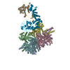
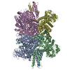
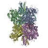
+ Open data
- Basic information
| Entry | Database: PDB / ID: 8kgy | |||||||||||||||||||||||||||||||||
|---|---|---|---|---|---|---|---|---|---|---|---|---|---|---|---|---|---|---|---|---|---|---|---|---|---|---|---|---|---|---|---|---|---|---|
| Title | Human glutamate dehydrogenase I | |||||||||||||||||||||||||||||||||
 Components Components | Glutamate dehydrogenase 1, mitochondrial | |||||||||||||||||||||||||||||||||
 Keywords Keywords | OXIDOREDUCTASE / dehydrogenase | |||||||||||||||||||||||||||||||||
| Function / homology |  Function and homology information Function and homology informationglutamate dehydrogenase [NAD(P)+] activity / L-leucine binding / tricarboxylic acid metabolic process / glutamate dehydrogenase [NAD(P)+] / glutamate biosynthetic process / glutamate dehydrogenase (NAD+) activity / glutamate dehydrogenase (NADP+) activity / Glutamate and glutamine metabolism / L-glutamate catabolic process / glutamine metabolic process ...glutamate dehydrogenase [NAD(P)+] activity / L-leucine binding / tricarboxylic acid metabolic process / glutamate dehydrogenase [NAD(P)+] / glutamate biosynthetic process / glutamate dehydrogenase (NAD+) activity / glutamate dehydrogenase (NADP+) activity / Glutamate and glutamine metabolism / L-glutamate catabolic process / glutamine metabolic process / NAD+ binding / Mitochondrial protein degradation / substantia nigra development / Transcriptional activation of mitochondrial biogenesis / positive regulation of insulin secretion / ADP binding / mitochondrial matrix / GTP binding / endoplasmic reticulum / protein homodimerization activity / mitochondrion / ATP binding / cytoplasm Similarity search - Function | |||||||||||||||||||||||||||||||||
| Biological species |  Homo sapiens (human) Homo sapiens (human) | |||||||||||||||||||||||||||||||||
| Method | ELECTRON MICROSCOPY / single particle reconstruction / cryo EM / Resolution: 2.59 Å | |||||||||||||||||||||||||||||||||
 Authors Authors | Su, M.-Y. | |||||||||||||||||||||||||||||||||
| Funding support |  China, 1items China, 1items
| |||||||||||||||||||||||||||||||||
 Citation Citation |  Journal: Structure / Year: 2023 Journal: Structure / Year: 2023Title: Cryo-EM structure of the KLHL22 E3 ligase bound to an oligomeric metabolic enzyme. Authors: Fei Teng / Yang Wang / Ming Liu / Shuyun Tian / Goran Stjepanovic / Ming-Yuan Su /  Abstract: CULLIN-RING ligases constitute the largest group of E3 ubiquitin ligases. While some CULLIN family members recruit adapters before engaging further with different substrate receptors, homo-dimeric ...CULLIN-RING ligases constitute the largest group of E3 ubiquitin ligases. While some CULLIN family members recruit adapters before engaging further with different substrate receptors, homo-dimeric BTB-Kelch family proteins combine adapter and substrate receptor into a single polypeptide for the CULLIN3 family. However, the entire structural assembly and molecular details have not been elucidated to date. Here, we present a cryo-EM structure of the CULLIN3 in complex with Kelch-like protein 22 (KLHL22) and a mitochondrial glutamate dehydrogenase complex I (GDH1) at 3.06 Å resolution. The structure adopts a W-shaped architecture formed by E3 ligase dimers. Three CULLIN3 dimers were found to be dynamically associated with a single GDH1 hexamer. CULLIN3 ligase mediated the polyubiquitination of GDH1 in vitro. Together, these results enabled the establishment of a structural model for understanding the complete assembly of BTB-Kelch proteins with CULLIN3 and how together they recognize oligomeric substrates and target them for ubiquitination. | |||||||||||||||||||||||||||||||||
| History |
|
- Structure visualization
| Structure viewer | Molecule:  Molmil Molmil Jmol/JSmol Jmol/JSmol |
|---|
- Downloads & links
- Download
| PDBx/mmCIF format |  8kgy.cif.gz 8kgy.cif.gz | 477.8 KB | Display |  PDBx/mmCIF format PDBx/mmCIF format |
|---|---|---|---|---|
| PDB format |  pdb8kgy.ent.gz pdb8kgy.ent.gz | 392.7 KB | Display |  PDB format PDB format |
| PDBx/mmJSON format |  8kgy.json.gz 8kgy.json.gz | Tree view |  PDBx/mmJSON format PDBx/mmJSON format | |
| Others |  Other downloads Other downloads |
-Validation report
| Summary document |  8kgy_validation.pdf.gz 8kgy_validation.pdf.gz | 381.8 KB | Display |  wwPDB validaton report wwPDB validaton report |
|---|---|---|---|---|
| Full document |  8kgy_full_validation.pdf.gz 8kgy_full_validation.pdf.gz | 402.3 KB | Display | |
| Data in XML |  8kgy_validation.xml.gz 8kgy_validation.xml.gz | 52.4 KB | Display | |
| Data in CIF |  8kgy_validation.cif.gz 8kgy_validation.cif.gz | 79.8 KB | Display | |
| Arichive directory |  https://data.pdbj.org/pub/pdb/validation_reports/kg/8kgy https://data.pdbj.org/pub/pdb/validation_reports/kg/8kgy ftp://data.pdbj.org/pub/pdb/validation_reports/kg/8kgy ftp://data.pdbj.org/pub/pdb/validation_reports/kg/8kgy | HTTPS FTP |
-Related structure data
| Related structure data |  37235MC  8khpC  8w4jC M: map data used to model this data C: citing same article ( |
|---|---|
| Similar structure data | Similarity search - Function & homology  F&H Search F&H Search |
- Links
Links
- Assembly
| Deposited unit | 
|
|---|---|
| 1 |
|
- Components
| #1: Protein | Mass: 61480.746 Da / Num. of mol.: 6 / Source method: isolated from a natural source / Source: (natural)  Homo sapiens (human) / Cell line: HEK Expi293F Homo sapiens (human) / Cell line: HEK Expi293FReferences: UniProt: P00367, glutamate dehydrogenase [NAD(P)+] Has protein modification | N | |
|---|
-Experimental details
-Experiment
| Experiment | Method: ELECTRON MICROSCOPY |
|---|---|
| EM experiment | Aggregation state: PARTICLE / 3D reconstruction method: single particle reconstruction |
- Sample preparation
| Component | Name: human glutamate dehydrogenase I / Type: COMPLEX / Entity ID: all / Source: NATURAL |
|---|---|
| Molecular weight | Units: MEGADALTONS / Experimental value: NO |
| Source (natural) | Organism:  Homo sapiens (human) Homo sapiens (human) |
| Buffer solution | pH: 7.4 Details: 20 mM HEPES pH 7.4, 200 mM NaCl, 2 mM MgCl2 and 0.5 mM TCEP |
| Specimen | Conc.: 0.6 mg/ml / Embedding applied: NO / Shadowing applied: NO / Staining applied: NO / Vitrification applied: YES |
| Specimen support | Grid material: GOLD / Grid mesh size: 300 divisions/in. / Grid type: UltrAuFoil R1.2/1.3 |
| Vitrification | Instrument: FEI VITROBOT MARK IV / Cryogen name: ETHANE / Humidity: 100 % / Chamber temperature: 277 K |
- Electron microscopy imaging
| Experimental equipment |  Model: Titan Krios / Image courtesy: FEI Company |
|---|---|
| Microscopy | Model: FEI TITAN KRIOS |
| Electron gun | Electron source:  FIELD EMISSION GUN / Accelerating voltage: 300 kV / Illumination mode: SPOT SCAN FIELD EMISSION GUN / Accelerating voltage: 300 kV / Illumination mode: SPOT SCAN |
| Electron lens | Mode: BRIGHT FIELD / Nominal defocus max: 1900 nm / Nominal defocus min: 1100 nm / Alignment procedure: COMA FREE |
| Specimen holder | Cryogen: NITROGEN |
| Image recording | Electron dose: 1.072 e/Å2 / Film or detector model: GATAN K3 (6k x 4k) |
- Processing
| EM software |
| ||||||||||||||||||||||||
|---|---|---|---|---|---|---|---|---|---|---|---|---|---|---|---|---|---|---|---|---|---|---|---|---|---|
| CTF correction | Type: PHASE FLIPPING AND AMPLITUDE CORRECTION | ||||||||||||||||||||||||
| Symmetry | Point symmetry: D3 (2x3 fold dihedral) | ||||||||||||||||||||||||
| 3D reconstruction | Resolution: 2.59 Å / Resolution method: FSC 0.143 CUT-OFF / Num. of particles: 79181 / Symmetry type: POINT | ||||||||||||||||||||||||
| Atomic model building | Protocol: RIGID BODY FIT / Space: REAL | ||||||||||||||||||||||||
| Atomic model building | PDB-ID: 1L1F Accession code: 1L1F / Source name: PDB / Type: experimental model | ||||||||||||||||||||||||
| Refine LS restraints |
|
 Movie
Movie Controller


 PDBj
PDBj
