 Movie
Movie Controller


+ Open data

- Basic information
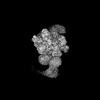
| Entry | Database: PDB / ID: 8khp | ||||||||||||||||||||||||
|---|---|---|---|---|---|---|---|---|---|---|---|---|---|---|---|---|---|---|---|---|---|---|---|---|---|
| Title | CULLIN3-KLHL22-RBX1 E3 ligase | ||||||||||||||||||||||||
 Components Components |
| ||||||||||||||||||||||||
 Keywords Keywords | STRUCTURAL PROTEIN / E3 ligase | ||||||||||||||||||||||||
| Function / homology |  Function and homology information Function and homology informationpositive regulation of mitotic cell cycle phase transition / POZ domain binding / polar microtubule / regulation protein catabolic process at postsynapse / anaphase-promoting complex-dependent catabolic process / nuclear protein quality control by the ubiquitin-proteasome system / COPII vesicle coat assembly / cell projection organization / cellular response to L-leucine / negative regulation of beige fat cell differentiation ...positive regulation of mitotic cell cycle phase transition / POZ domain binding / polar microtubule / regulation protein catabolic process at postsynapse / anaphase-promoting complex-dependent catabolic process / nuclear protein quality control by the ubiquitin-proteasome system / COPII vesicle coat assembly / cell projection organization / cellular response to L-leucine / negative regulation of beige fat cell differentiation / embryonic cleavage / RHOBTB3 ATPase cycle / positive regulation of T cell mediated immune response to tumor cell / cullin-RING-type E3 NEDD8 transferase / NEDD8 transferase activity / cullin-RING ubiquitin ligase complex / stem cell division / Cul7-RING ubiquitin ligase complex / cellular response to chemical stress / Loss of Function of FBXW7 in Cancer and NOTCH1 Signaling / positive regulation of mitotic metaphase/anaphase transition / positive regulation of protein autoubiquitination / Notch binding / RNA polymerase II transcription initiation surveillance / protein neddylation / negative regulation of Rho protein signal transduction / NEDD8 ligase activity / negative regulation of response to oxidative stress / RHOBTB1 GTPase cycle / protein K27-linked ubiquitination / VCB complex / Cul5-RING ubiquitin ligase complex / ubiquitin-ubiquitin ligase activity / ubiquitin-dependent protein catabolic process via the C-end degron rule pathway / mitotic spindle assembly checkpoint signaling / SCF ubiquitin ligase complex / Cul2-RING ubiquitin ligase complex / stress fiber assembly / Cul3-RING ubiquitin ligase complex / negative regulation of type I interferon production / positive regulation of cytokinesis / SCF-dependent proteasomal ubiquitin-dependent protein catabolic process / Prolactin receptor signaling / negative regulation of mitophagy / Cul4A-RING E3 ubiquitin ligase complex / intercellular bridge / Cul4-RING E3 ubiquitin ligase complex / Cul4B-RING E3 ubiquitin ligase complex / ubiquitin ligase complex scaffold activity / mitotic sister chromatid segregation / protein monoubiquitination / mitotic metaphase chromosome alignment / cullin family protein binding / endoplasmic reticulum to Golgi vesicle-mediated transport / sperm flagellum / RHOBTB2 GTPase cycle / SPOP-mediated proteasomal degradation of PD-L1(CD274) / protein autoubiquitination / ubiquitin-like ligase-substrate adaptor activity / site of DNA damage / signal transduction in response to DNA damage / Nuclear events stimulated by ALK signaling in cancer / protein K48-linked ubiquitination / kidney development / negative regulation of insulin receptor signaling pathway / 14-3-3 protein binding / regulation of cellular response to insulin stimulus / positive regulation of TORC1 signaling / transcription-coupled nucleotide-excision repair / post-translational protein modification / intrinsic apoptotic signaling pathway / cyclin binding / negative regulation of autophagy / integrin-mediated signaling pathway / positive regulation of protein ubiquitination / negative regulation of canonical NF-kappaB signal transduction / cellular response to amino acid stimulus / Regulation of BACH1 activity / T cell activation / G1/S transition of mitotic cell cycle / negative regulation of canonical Wnt signaling pathway / protein destabilization / Degradation of DVL / Degradation of CRY and PER proteins / Degradation of GLI1 by the proteasome / RING-type E3 ubiquitin transferase / Recognition of DNA damage by PCNA-containing replication complex / GSK3B and BTRC:CUL1-mediated-degradation of NFE2L2 / Negative regulation of NOTCH4 signaling / Hedgehog 'on' state / Vif-mediated degradation of APOBEC3G / FBXL7 down-regulates AURKA during mitotic entry and in early mitosis / Degradation of GLI2 by the proteasome / GLI3 is processed to GLI3R by the proteasome / Ubiquitin-Mediated Degradation of Phosphorylated Cdc25A / Evasion by RSV of host interferon responses / NOTCH1 Intracellular Domain Regulates Transcription / Degradation of beta-catenin by the destruction complex / DNA Damage Recognition in GG-NER / Oxygen-dependent proline hydroxylation of Hypoxia-inducible Factor Alpha Similarity search - Function | ||||||||||||||||||||||||
| Biological species |  Homo sapiens (human) Homo sapiens (human) | ||||||||||||||||||||||||
| Method | ELECTRON MICROSCOPY / single particle reconstruction / cryo EM / Resolution: 3.67 Å | ||||||||||||||||||||||||
 Authors Authors | Su, M.-Y. / Su, M.-Y. | ||||||||||||||||||||||||
| Funding support |  China, 1items China, 1items
| ||||||||||||||||||||||||
 Citation Citation | Journal: Structure / Year: 2023 Title: Cryo-EM structure of the KLHL22 E3 ligase bound to an oligomeric metabolic enzyme. Authors: Fei Teng / Yang Wang / Ming Liu / Shuyun Tian / Goran Stjepanovic / Ming-Yuan Su / Abstract: CULLIN-RING ligases constitute the largest group of E3 ubiquitin ligases. While some CULLIN family members recruit adapters before engaging further with different substrate receptors, homo-dimeric ...CULLIN-RING ligases constitute the largest group of E3 ubiquitin ligases. While some CULLIN family members recruit adapters before engaging further with different substrate receptors, homo-dimeric BTB-Kelch family proteins combine adapter and substrate receptor into a single polypeptide for the CULLIN3 family. However, the entire structural assembly and molecular details have not been elucidated to date. Here, we present a cryo-EM structure of the CULLIN3 in complex with Kelch-like protein 22 (KLHL22) and a mitochondrial glutamate dehydrogenase complex I (GDH1) at 3.06 Å resolution. The structure adopts a W-shaped architecture formed by E3 ligase dimers. Three CULLIN3 dimers were found to be dynamically associated with a single GDH1 hexamer. CULLIN3 ligase mediated the polyubiquitination of GDH1 in vitro. Together, these results enabled the establishment of a structural model for understanding the complete assembly of BTB-Kelch proteins with CULLIN3 and how together they recognize oligomeric substrates and target them for ubiquitination. | ||||||||||||||||||||||||
| History |
|
- Structure visualization
| Structure viewer | Molecule: MolmilJmol/JSmol |
|---|
- Downloads & links
-Download
| PDBx/mmCIF format | 8khp.cif.gz | 411.7 KB | Display | PDBx/mmCIF format |
|---|---|---|---|---|
| PDB format | pdb8khp.ent.gz | 305.2 KB | Display | PDB format |
| PDBx/mmJSON format | 8khp.json.gz | Tree view | PDBx/mmJSON format | |
| Others |  Other downloads Other downloads |
-Validation report
| Arichive directory | https://data.pdbj.org/pub/pdb/validation_reports/kh/8khpftp://data.pdbj.org/pub/pdb/validation_reports/kh/8khp | HTTPS FTP |
|---|
-Related structure data
| Related structure data | 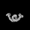 37247MC 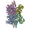 8kgyC 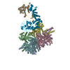 8w4jC C: citing same article ( M: map data used to model this data |
|---|---|
| Similar structure data |
-Links
 PDBj
PDBj


- Assembly
| Deposited unit | 
|
|---|---|
| 1 |
|
-Components
| #1: Protein | Mass: 71744.594 Da / Num. of mol.: 2 Source method: isolated from a genetically manipulated source Source: (gene. exp.) Homo sapiens (human) / Gene: KLHL22 / Production host: Homo sapiens (human) / References: UniProt: Q53GT1#2: Protein | Mass: 89063.328 Da / Num. of mol.: 2 Source method: isolated from a genetically manipulated source Source: (gene. exp.) Homo sapiens (human) / Gene: CUL3, KIAA0617 / Production host: Homo sapiens (human) / References: UniProt: Q13618#3: Protein | Mass: 12289.977 Da / Num. of mol.: 2 Source method: isolated from a genetically manipulated source Source: (gene. exp.) Homo sapiens (human) / Gene: RBX1, RNF75, ROC1 / Production host: Homo sapiens (human)References: UniProt: P62877, RING-type E3 ubiquitin transferase, cullin-RING-type E3 NEDD8 transferase Has protein modification | N | |
|---|
-Experimental details
-Experiment
| Experiment | Method: ELECTRON MICROSCOPY |
|---|---|
| EM experiment | Aggregation state: PARTICLE / 3D reconstruction method: single particle reconstruction |
- Sample preparation
| Component | Name: CULLIN3-KLHL22-RBX1 E3 ligase / Type: COMPLEX / Entity ID: all / Source: RECOMBINANT |
|---|---|
| Molecular weight | Experimental value: NO |
| Source (natural) | Organism: Homo sapiens (human) |
| Source (recombinant) | Organism: Homo sapiens (human) |
| Buffer solution | pH: 7.4 |
| Specimen | Embedding applied: NO / Shadowing applied: NO / Staining applied: NO / Vitrification applied: YES |
| Vitrification | Cryogen name: ETHANE |
- Electron microscopy imaging
| Experimental equipment |  Model: Titan Krios / Image courtesy: FEI Company |
|---|---|
| Microscopy | Model: FEI TITAN KRIOS |
| Electron gun | Electron source:  FIELD EMISSION GUN / Accelerating voltage: 300 kV / Illumination mode: SPOT SCAN FIELD EMISSION GUN / Accelerating voltage: 300 kV / Illumination mode: SPOT SCAN |
| Electron lens | Mode: BRIGHT FIELD / Nominal defocus max: 1900 nm / Nominal defocus min: 1100 nm |
| Image recording | Electron dose: 1.07 e/Å2 / Film or detector model: GATAN K3 (6k x 4k) |
- Processing
| EM software | Name: PHENIX / Category: model refinement | ||||||||||||||||||||||||
|---|---|---|---|---|---|---|---|---|---|---|---|---|---|---|---|---|---|---|---|---|---|---|---|---|---|
| CTF correction | Type: NONE | ||||||||||||||||||||||||
| 3D reconstruction | Resolution: 3.67 Å / Resolution method: FSC 0.143 CUT-OFF / Num. of particles: 181834 / Symmetry type: POINT | ||||||||||||||||||||||||
| Refine LS restraints |
|
