 Movie
Movie Controller


+ Open data

- Basic information
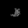
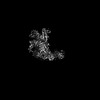
| Entry | Database: PDB / ID: 8jxg | ||||||
|---|---|---|---|---|---|---|---|
| Title | rat megalin RAP complex bodyB | ||||||
 Components Components |
| ||||||
 Keywords Keywords | ENDOCYTOSIS / endocytosis receptor | ||||||
| Function / homology |  Function and homology information Function and homology informationchemoattraction of axon / diol metabolic process / pulmonary artery morphogenesis / secondary heart field specification / positive regulation of oligodendrocyte progenitor proliferation / folate import across plasma membrane / ventricular compact myocardium morphogenesis / response to leptin / metal ion transport / vitamin D metabolic process ...chemoattraction of axon / diol metabolic process / pulmonary artery morphogenesis / secondary heart field specification / positive regulation of oligodendrocyte progenitor proliferation / folate import across plasma membrane / ventricular compact myocardium morphogenesis / response to leptin / metal ion transport / vitamin D metabolic process / cranial skeletal system development / coronary artery morphogenesis / neuron projection arborization / outflow tract septum morphogenesis / ventricular septum development / aorta development / forebrain development / vagina development / negative regulation of BMP signaling pathway / positive regulation of endocytosis / endocytic vesicle / axonal growth cone / clathrin-coated pit / receptor-mediated endocytosis / endosome lumen / kidney development / neural tube closure / brush border membrane / phosphatidylinositol 3-kinase/protein kinase B signal transduction / sensory perception of sound / SH3 domain binding / male gonad development / protein transport / protein-folding chaperone binding / gene expression / signaling receptor complex / cell population proliferation / external side of plasma membrane / calcium ion binding / dendrite / negative regulation of apoptotic process / endoplasmic reticulum / Golgi apparatus Similarity search - Function | ||||||
| Biological species |  | ||||||
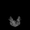
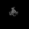
| Method | ELECTRON MICROSCOPY / single particle reconstruction / cryo EM / Resolution: 3.2 Å | ||||||
 Authors Authors | Goto, S. / Tsutsumi, A. / Lee, Y. / Hosojima, M. / Kabasawa, H. / Komochi, K. / Yun-san, L. / Nagatoshi, S. / Tsumoto, K. / Nishizawa, T. ...Goto, S. / Tsutsumi, A. / Lee, Y. / Hosojima, M. / Kabasawa, H. / Komochi, K. / Yun-san, L. / Nagatoshi, S. / Tsumoto, K. / Nishizawa, T. / Kikkawa, M. / Saito, A. | ||||||
| Funding support |  Japan, 1items Japan, 1items
| ||||||
 Citation Citation | Journal: Proc Natl Acad Sci U S A / Year: 2024 Title: Cryo-EM structures elucidate the multiligand receptor nature of megalin. Authors: Sawako Goto / Akihisa Tsutsumi / Yongchan Lee / Michihiro Hosojima / Hideyuki Kabasawa / Koichi Komochi / Satoru Nagatoishi / Kazuya Takemoto / Kouhei Tsumoto / Tomohiro Nishizawa / Masahide ...Authors: Sawako Goto / Akihisa Tsutsumi / Yongchan Lee / Michihiro Hosojima / Hideyuki Kabasawa / Koichi Komochi / Satoru Nagatoishi / Kazuya Takemoto / Kouhei Tsumoto / Tomohiro Nishizawa / Masahide Kikkawa / Akihiko Saito / Abstract: Megalin (low-density lipoprotein receptor-related protein 2) is a giant glycoprotein of about 600 kDa, mediating the endocytosis of more than 60 ligands, including those of proteins, peptides, and ...Megalin (low-density lipoprotein receptor-related protein 2) is a giant glycoprotein of about 600 kDa, mediating the endocytosis of more than 60 ligands, including those of proteins, peptides, and drug compounds [S. Goto, M. Hosojima, H. Kabasawa, A. Saito, , 106393 (2023)]. It is expressed predominantly in renal proximal tubule epithelial cells, as well as in the brain, lungs, eyes, inner ear, thyroid gland, and placenta. Megalin is also known to mediate the endocytosis of toxic compounds, particularly those that cause renal and hearing disorders [Y. Hori , , 1783-1791 (2017)]. Genetic megalin deficiency causes Donnai-Barrow syndrome/facio-oculo-acoustico-renal syndrome in humans. However, it is not known how megalin interacts with such a wide variety of ligands and plays pathological roles in various organs. In this study, we elucidated the dimeric architecture of megalin, purified from rat kidneys, using cryoelectron microscopy. The maps revealed the densities of endogenous ligands bound to various regions throughout the dimer, elucidating the multiligand receptor nature of megalin. We also determined the structure of megalin in complex with receptor-associated protein, a molecular chaperone for megalin. The results will facilitate further studies on the pathophysiology of megalin-dependent multiligand endocytic pathways in multiple organs and will also be useful for the development of megalin-targeted drugs for renal and hearing disorders, Alzheimer's disease [B. V. Zlokovic , , 4229-4234 (1996)], and other illnesses. | ||||||
| History |
|
- Structure visualization
| Structure viewer | Molecule: MolmilJmol/JSmol |
|---|
- Downloads & links
-Download
| PDBx/mmCIF format | 8jxg.cif.gz | 338.9 KB | Display | PDBx/mmCIF format |
|---|---|---|---|---|
| PDB format | pdb8jxg.ent.gz | 226 KB | Display | PDB format |
| PDBx/mmJSON format | 8jxg.json.gz | Tree view | PDBx/mmJSON format | |
| Others |  Other downloads Other downloads |
-Validation report
| Arichive directory | https://data.pdbj.org/pub/pdb/validation_reports/jx/8jxgftp://data.pdbj.org/pub/pdb/validation_reports/jx/8jxg | HTTPS FTP |
|---|
-Related structure data
| Related structure data | 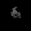 36700MC 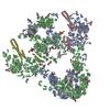 8jutC 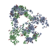 8juuC 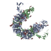 8jx8C 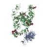 8jx9C 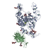 8jxaC 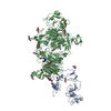 8jxbC 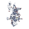 8jxcC 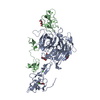 8jxdC 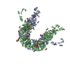 8jxeC 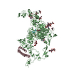 8jxfC 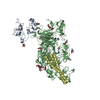 8jxhC 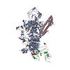 8jxiC 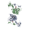 8jxjC C: citing same article ( M: map data used to model this data |
|---|---|
| Similar structure data |


-Links
 PDBj
PDBj


- Assembly
| Deposited unit | 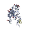
|
|---|---|
| 1 |
|
-Components
-Protein , 2 types, 2 molecules DA
| #1: Protein | Mass: 38610.309 Da / Num. of mol.: 1 Source method: isolated from a genetically manipulated source Source: (gene. exp.)  |
|---|---|
| #2: Protein | Mass: 519871.438 Da / Num. of mol.: 1 / Source method: isolated from a natural source / Source: (natural) |
-Protein/peptide / Non-polymers , 2 types, 22 molecules M
| #3: Protein/peptide | Mass: 472.537 Da / Num. of mol.: 1 / Source method: isolated from a natural source Details: Authors could not experimentally confirm this sequence, but the sequence should be a part of molecule 2. Therefore the residue numbers and chain ID are meaningless for molecule 3. Source: (natural) |
|---|---|
| #8: Chemical | ChemComp-CA /  Mass: 40.078 Da / Num. of mol.: 21 / Source method: obtained synthetically / Formula: Ca Mass: 40.078 Da / Num. of mol.: 21 / Source method: obtained synthetically / Formula: Ca |
-Sugars , 4 types, 13 molecules 

| #4: Polysaccharide | beta-D-mannopyranose-(1-4)-2-acetamido-2-deoxy-beta-D-glucopyranose-(1-4)-2-acetamido-2-deoxy-beta- ...beta-D-mannopyranose-(1-4)-2-acetamido-2-deoxy-beta-D-glucopyranose-(1-4)-2-acetamido-2-deoxy-beta-D-glucopyranose Source method: isolated from a genetically manipulated source #5: Polysaccharide | Source method: isolated from a genetically manipulated source #6: Sugar |  Type: D-saccharide, beta linking / Mass: 221.208 Da / Num. of mol.: 2 / Source method: obtained synthetically / Formula: C8H15NO6 Type: D-saccharide, beta linking / Mass: 221.208 Da / Num. of mol.: 2 / Source method: obtained synthetically / Formula: C8H15NO6#7: Sugar | ChemComp-A2G /  Type: D-saccharide, alpha linking / Mass: 221.208 Da / Num. of mol.: 4 / Source method: obtained synthetically / Formula: C8H15NO6 Type: D-saccharide, alpha linking / Mass: 221.208 Da / Num. of mol.: 4 / Source method: obtained synthetically / Formula: C8H15NO6 |
|---|
-Details
| Has ligand of interest | N |
|---|---|
| Has protein modification | Y |
-Experimental details
-Experiment
| Experiment | Method: ELECTRON MICROSCOPY |
|---|---|
| EM experiment | Aggregation state: PARTICLE / 3D reconstruction method: single particle reconstruction |
- Sample preparation
| Component |
| ||||||||||||||||||||||||
|---|---|---|---|---|---|---|---|---|---|---|---|---|---|---|---|---|---|---|---|---|---|---|---|---|---|
| Source (natural) |
| ||||||||||||||||||||||||
| Source (recombinant) | Organism: | ||||||||||||||||||||||||
| Buffer solution | pH: 7.4 | ||||||||||||||||||||||||
| Specimen | Embedding applied: NO / Shadowing applied: NO / Staining applied: NO / Vitrification applied: YES | ||||||||||||||||||||||||
| Vitrification | Instrument: FEI VITROBOT MARK IV / Cryogen name: ETHANE |
- Electron microscopy imaging
| Experimental equipment |  Model: Titan Krios / Image courtesy: FEI Company |
|---|---|
| Microscopy | Model: FEI TITAN KRIOS |
| Electron gun | Electron source:  FIELD EMISSION GUN / Accelerating voltage: 300 kV / Illumination mode: FLOOD BEAM FIELD EMISSION GUN / Accelerating voltage: 300 kV / Illumination mode: FLOOD BEAM |
| Electron lens | Mode: BRIGHT FIELD / Nominal defocus max: 1600 nm / Nominal defocus min: 600 nm |
| Image recording | Electron dose: 50 e/Å2 / Film or detector model: GATAN K3 BIOQUANTUM (6k x 4k) |
- Processing
| EM software |
| ||||||||||||||||||||||||||||||||||||||||||||
|---|---|---|---|---|---|---|---|---|---|---|---|---|---|---|---|---|---|---|---|---|---|---|---|---|---|---|---|---|---|---|---|---|---|---|---|---|---|---|---|---|---|---|---|---|---|
| CTF correction | Type: PHASE FLIPPING ONLY | ||||||||||||||||||||||||||||||||||||||||||||
| Symmetry | Point symmetry: C1 (asymmetric) | ||||||||||||||||||||||||||||||||||||||||||||
| 3D reconstruction | Resolution: 3.2 Å / Resolution method: FSC 0.143 CUT-OFF / Num. of particles: 67775 / Algorithm: BACK PROJECTION / Symmetry type: POINT | ||||||||||||||||||||||||||||||||||||||||||||
| Atomic model building | Source name: AlphaFold / Type: in silico model |
