 Movie
Movie Controller

[English] 日本語
 Yorodumi
- PDB-8jgn: Cryo-EM structure of alpha/beta hydrolase DANGEROUS MIX 3 (DM3) f... -
+ Open data

- Basic information
| Entry | Database: PDB / ID: 8jgn | ||||||
|---|---|---|---|---|---|---|---|
| Title | Cryo-EM structure of alpha/beta hydrolase DANGEROUS MIX 3 (DM3) forms compartmentalized hexamer structure | ||||||
 Components Components | AT3g61540/F2A19_140 | ||||||
 Keywords Keywords | HYDROLASE / Complex / immune related alpha/beta hydrolase / PROLYL AMINOPEPTIDASE 2 | ||||||
| Function / homology |  Function and homology information Function and homology information | ||||||
| Biological species |  | ||||||
| Method | ELECTRON MICROSCOPY / single particle reconstruction / cryo EM / Resolution: 3.39 Å | ||||||
 Authors Authors | Kim, G. / Song, J.J. | ||||||
| Funding support |  Korea, Republic Of, 1items Korea, Republic Of, 1items
| ||||||
 Citation Citation | Journal: Mol.Cell / Year: 2025 Title: Structure determinants of DANGEROUS MIX 3, an alpha/beta hydrolase, for triggering NLR-mediated genetic incompatibility in plants Authors: Kim, G. / Song, J.J. | ||||||
| History |
|
- Structure visualization
| Structure viewer | Molecule: MolmilJmol/JSmol |
|---|
- Downloads & links
-Download
| PDBx/mmCIF format | 8jgn.cif.gz | 456.3 KB | Display | PDBx/mmCIF format |
|---|---|---|---|---|
| PDB format | pdb8jgn.ent.gz | 382.1 KB | Display | PDB format |
| PDBx/mmJSON format | 8jgn.json.gz | Tree view | PDBx/mmJSON format | |
| Others |  Other downloads Other downloads |
-Validation report
| Arichive directory | https://data.pdbj.org/pub/pdb/validation_reports/jg/8jgnftp://data.pdbj.org/pub/pdb/validation_reports/jg/8jgn | HTTPS FTP |
|---|
-Related structure data
| Related structure data |  36240MC 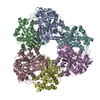 8jgmC M: map data used to model this data C: citing same article ( |
|---|---|
| Similar structure data |

-Links
 PDBj
PDBj


- Assembly
| Deposited unit | 
|
|---|---|
| 1 |
|
-Components
| #1: Protein | Mass: 50432.719 Da / Num. of mol.: 6 Source method: isolated from a genetically manipulated source Source: (gene. exp.)  Has protein modification | N | |
|---|
-Experimental details
-Experiment
| Experiment | Method: ELECTRON MICROSCOPY |
|---|---|
| EM experiment | Aggregation state: PARTICLE / 3D reconstruction method: single particle reconstruction |
- Sample preparation
| Component | Name: Homohexamer complex of DM3 from Columbia accession (Col-0) Type: COMPLEX / Entity ID: all / Source: RECOMBINANT | |||||||||||||||
|---|---|---|---|---|---|---|---|---|---|---|---|---|---|---|---|---|
| Molecular weight | Experimental value: NO | |||||||||||||||
| Source (natural) | Organism: | |||||||||||||||
| Source (recombinant) | Organism: | |||||||||||||||
| Buffer solution | pH: 8 | |||||||||||||||
| Buffer component |
| |||||||||||||||
| Specimen | Conc.: 0.2 mg/ml / Embedding applied: NO / Shadowing applied: NO / Staining applied: NO / Vitrification applied: YES | |||||||||||||||
| Specimen support | Details: After glow discharge, graphene oxide was treated onto the grid Grid material: COPPER / Grid mesh size: 300 divisions/in. / Grid type: Quantifoil R1.2/1.3 | |||||||||||||||
| Vitrification | Instrument: FEI VITROBOT MARK IV / Cryogen name: ETHANE / Humidity: 100 % / Chamber temperature: 277 K |
- Electron microscopy imaging
| Microscopy | Model: TFS GLACIOS |
|---|---|
| Electron gun | Electron source:  FIELD EMISSION GUN / Accelerating voltage: 200 kV / Illumination mode: OTHER FIELD EMISSION GUN / Accelerating voltage: 200 kV / Illumination mode: OTHER |
| Electron lens | Mode: BRIGHT FIELD / Nominal defocus max: 2000 nm / Nominal defocus min: 800 nm / Cs: 2.7 mm |
| Specimen holder | Cryogen: NITROGEN |
| Image recording | Electron dose: 40 e/Å2 / Detector mode: COUNTING / Film or detector model: FEI FALCON III (4k x 4k) / Num. of grids imaged: 1 |
- Processing
| CTF correction | Type: PHASE FLIPPING AND AMPLITUDE CORRECTION |
|---|---|
| Symmetry | Point symmetry: C1 (asymmetric) |
| 3D reconstruction | Resolution: 3.39 Å / Resolution method: FSC 0.143 CUT-OFF / Num. of particles: 330509 / Symmetry type: POINT |
