 Movie
Movie Controller

[English] 日本語
 Yorodumi
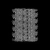
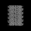
- PDB-8ixf: GMPCPP-Alpha4A/Beta2A-microtubule decorated with kinesin non-seam... -
+ Open data

- Basic information
| Entry | Database: PDB / ID: 8ixf | ||||||
|---|---|---|---|---|---|---|---|
| Title | GMPCPP-Alpha4A/Beta2A-microtubule decorated with kinesin non-seam region | ||||||
 Components Components |
| ||||||
 Keywords Keywords | STRUCTURAL PROTEIN / microtubule / tubulin isotype / cryo-EM structure | ||||||
| Function / homology |  Function and homology information Function and homology informationMicrotubule-dependent trafficking of connexons from Golgi to the plasma membrane / Cilium Assembly / Sealing of the nuclear envelope (NE) by ESCRT-III / Carboxyterminal post-translational modifications of tubulin / Intraflagellar transport / COPI-independent Golgi-to-ER retrograde traffic / regulation of modification of synapse structure, modulating synaptic transmission / HSP90 chaperone cycle for steroid hormone receptors (SHR) in the presence of ligand / plus-end-directed vesicle transport along microtubule / cytoplasm organization ...Microtubule-dependent trafficking of connexons from Golgi to the plasma membrane / Cilium Assembly / Sealing of the nuclear envelope (NE) by ESCRT-III / Carboxyterminal post-translational modifications of tubulin / Intraflagellar transport / COPI-independent Golgi-to-ER retrograde traffic / regulation of modification of synapse structure, modulating synaptic transmission / HSP90 chaperone cycle for steroid hormone receptors (SHR) in the presence of ligand / plus-end-directed vesicle transport along microtubule / cytoplasm organization / cytolytic granule membrane / anterograde dendritic transport of neurotransmitter receptor complex / COPI-mediated anterograde transport / Aggrephagy / Kinesins / anterograde neuronal dense core vesicle transport / Mitotic Prometaphase / EML4 and NUDC in mitotic spindle formation / Resolution of Sister Chromatid Cohesion / PKR-mediated signaling / mitocytosis / retrograde neuronal dense core vesicle transport / The role of GTSE1 in G2/M progression after G2 checkpoint / RHO GTPases activate IQGAPs / Recycling pathway of L1 / anterograde axonal protein transport / COPI-dependent Golgi-to-ER retrograde traffic / RHO GTPases Activate Formins / Separation of Sister Chromatids / Hedgehog 'off' state / Loss of Nlp from mitotic centrosomes / Recruitment of mitotic centrosome proteins and complexes / Loss of proteins required for interphase microtubule organization from the centrosome / Recruitment of NuMA to mitotic centrosomes / Anchoring of the basal body to the plasma membrane / AURKA Activation by TPX2 / Platelet degranulation / Regulation of PLK1 Activity at G2/M Transition / ciliary rootlet / lysosome localization / positive regulation of potassium ion transport / MHC class II antigen presentation / plus-end-directed microtubule motor activity / Kinesins / vesicle transport along microtubule / RHO GTPases activate KTN1 / kinesin complex / microtubule motor activity / mitochondrion transport along microtubule / centrosome localization / COPI-dependent Golgi-to-ER retrograde traffic / natural killer cell mediated cytotoxicity / stress granule disassembly / microtubule-based movement / Insulin processing / synaptic vesicle transport / postsynaptic cytosol / microtubule-based process / intercellular bridge / phagocytic vesicle / axon cytoplasm / MHC class II antigen presentation / dendrite cytoplasm / axon guidance / positive regulation of synaptic transmission, GABAergic / positive regulation of protein localization to plasma membrane / regulation of membrane potential / cerebral cortex development / structural constituent of cytoskeleton / cellular response to type II interferon / centriolar satellite / mitotic spindle / Signaling by ALK fusions and activated point mutants / nuclear membrane / microtubule binding / vesicle / Hydrolases; Acting on acid anhydrides; Acting on GTP to facilitate cellular and subcellular movement / microtubule / cilium / cadherin binding / hydrolase activity / GTPase activity / protein kinase binding / GTP binding / protein-containing complex binding / perinuclear region of cytoplasm / enzyme binding / ATP hydrolysis activity / mitochondrion / ATP binding / metal ion binding / identical protein binding / membrane / cytosol / cytoplasm Similarity search - Function | ||||||
| Biological species |   Homo sapiens (human) Homo sapiens (human) | ||||||
| Method | ELECTRON MICROSCOPY / single particle reconstruction / cryo EM / Resolution: 4.4 Å | ||||||
 Authors Authors | Zheng, W. / Zhao, Q.Y. / Diao, L. / Bao, L. / Cong, Y. | ||||||
| Funding support |  China, 1items China, 1items
| ||||||
 Citation Citation | Journal: Acta Biochim Biophys Sin (Shanghai) / Year: 2023 Title: Cryo-EM of α-tubulin isotype-containing microtubules revealed a contracted structure of α4A/β2A microtubules. Authors: Lei Diao / Wei Zheng / Qiaoyu Zhao / Mingyi Liu / Zhenglin Fu / Xu Zhang / Lan Bao / Yao Cong / Abstract: Microtubules are hollow α/β-tubulin heterodimeric polymers that play critical roles in cells. In vertebrates, both α- and β-tubulins have multiple isotypes encoded by different genes, which are ...Microtubules are hollow α/β-tubulin heterodimeric polymers that play critical roles in cells. In vertebrates, both α- and β-tubulins have multiple isotypes encoded by different genes, which are intrinsic factors in regulating microtubule functions. However, the structures of microtubules composed of different tubulin isotypes, especially α-tubulin isotypes, remain largely unknown. Here, we purify recombinant tubulin heterodimers composed of different mouse α-tubulin isotypes, including α1A, α1C and α4A, with the β-tubulin isotype β2A. We further assemble and determine the cryo-electron microscopy (cryo-EM) structures of α1A/β2A, α1C/β2A, and α4A/β2A microtubules. Our structural analysis demonstrates that α4A/β2A microtubules exhibit longitudinal contraction between tubulin interdimers compared with α1A/β2A and α1C/β2A microtubules. Collectively, our findings reveal that α-tubulin isotype composition can tune microtubule structures, and also provide evidence for the "tubulin code" hypothesis. | ||||||
| History |
|
- Structure visualization
| Structure viewer | Molecule: MolmilJmol/JSmol |
|---|
- Downloads & links
-Download
| PDBx/mmCIF format | 8ixf.cif.gz | 1.8 MB | Display | PDBx/mmCIF format |
|---|---|---|---|---|
| PDB format | pdb8ixf.ent.gz | 1.5 MB | Display | PDB format |
| PDBx/mmJSON format | 8ixf.json.gz | Tree view | PDBx/mmJSON format | |
| Others |  Other downloads Other downloads |
-Validation report
| Arichive directory | https://data.pdbj.org/pub/pdb/validation_reports/ix/8ixfftp://data.pdbj.org/pub/pdb/validation_reports/ix/8ixf | HTTPS FTP |
|---|
-Related structure data
| Related structure data | 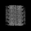 35792MC  8ixaC  8ixbC  8ixdC  8ixeC  8ixgC M: map data used to model this data C: citing same article ( |
|---|---|
| Similar structure data |
-Links
 PDBj
PDBj


- Assembly
| Deposited unit | 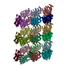
|
|---|---|
| 1 |
|
-Components
-Protein , 3 types, 27 molecules IABCDEFGHQJKLMNOPRYSTUVWXZa
| #1: Protein | Mass: 50806.109 Da / Num. of mol.: 9 Source method: isolated from a genetically manipulated source Details: Author stated: the data processing is a non-standard procedure for microtubule-type of helical reconstruction with a seam, and it was performed around 2017 to 2019. We followed the procedure ...Details: Author stated: the data processing is a non-standard procedure for microtubule-type of helical reconstruction with a seam, and it was performed around 2017 to 2019. We followed the procedure developed by Dr. Rui Zhang (R. Zhang, Cell, 2015, doi: 10.1016/j.cell.2015.07.012), with all the scripts provided by him. In this way, the generated half-maps have an applied circular mask but without touching the structural outer boundaries. Source: (gene. exp.)  Insecta environmental sample (insect) Insecta environmental sample (insect)References: UniProt: P68368, Hydrolases; Acting on acid anhydrides; Acting on GTP to facilitate cellular and subcellular movement #2: Protein | Mass: 51150.961 Da / Num. of mol.: 9 Source method: isolated from a genetically manipulated source Source: (gene. exp.) Insecta environmental sample (insect) / References: UniProt: Q7TMM9#3: Protein | Mass: 41721.066 Da / Num. of mol.: 9 Source method: isolated from a genetically manipulated source Source: (gene. exp.) Homo sapiens (human) / Gene: KIF5B, KNS, KNS1 / Production host:  |
|---|
-Non-polymers , 3 types, 27 molecules 


| #4: Chemical | ChemComp-GTP /  Mass: 523.180 Da / Num. of mol.: 9 / Source method: obtained synthetically / Formula: C10H16N5O14P3 / Comment: GTP, energy-carrying molecule*YM Mass: 523.180 Da / Num. of mol.: 9 / Source method: obtained synthetically / Formula: C10H16N5O14P3 / Comment: GTP, energy-carrying molecule*YM#5: Chemical | ChemComp-G2P /  Mass: 521.208 Da / Num. of mol.: 9 / Source method: obtained synthetically / Formula: C11H18N5O13P3 / Comment: GMP-CPP, energy-carrying molecule analogue*YM Mass: 521.208 Da / Num. of mol.: 9 / Source method: obtained synthetically / Formula: C11H18N5O13P3 / Comment: GMP-CPP, energy-carrying molecule analogue*YM#6: Chemical | ChemComp-ATP /  Mass: 507.181 Da / Num. of mol.: 9 / Source method: obtained synthetically / Formula: C10H16N5O13P3 / Comment: ATP, energy-carrying molecule*YM Mass: 507.181 Da / Num. of mol.: 9 / Source method: obtained synthetically / Formula: C10H16N5O13P3 / Comment: ATP, energy-carrying molecule*YM |
|---|
-Details
| Has ligand of interest | N |
|---|
-Experimental details
-Experiment
| Experiment | Method: ELECTRON MICROSCOPY |
|---|---|
| EM experiment | Aggregation state: FILAMENT / 3D reconstruction method: single particle reconstruction |
- Sample preparation
| Component | Name: GMPCPP-Alpha4A/Beta2A-microtubule decorated with kinesin Type: COMPLEX / Entity ID: #1-#3 / Source: RECOMBINANT |
|---|---|
| Source (natural) | Organism: |
| Source (recombinant) | Organism: Insecta environmental sample (insect) |
| Buffer solution | pH: 6.9 |
| Specimen | Embedding applied: NO / Shadowing applied: NO / Staining applied: NO / Vitrification applied: YES |
| Vitrification | Cryogen name: ETHANE |
- Electron microscopy imaging
| Experimental equipment |  Model: Titan Krios / Image courtesy: FEI Company |
|---|---|
| Microscopy | Model: FEI TITAN KRIOS |
| Electron gun | Electron source:  FIELD EMISSION GUN / Accelerating voltage: 300 kV / Illumination mode: FLOOD BEAM FIELD EMISSION GUN / Accelerating voltage: 300 kV / Illumination mode: FLOOD BEAM |
| Electron lens | Mode: BRIGHT FIELD / Nominal defocus max: 1500 nm / Nominal defocus min: 800 nm |
| Image recording | Electron dose: 36 e/Å2 / Detector mode: COUNTING / Film or detector model: GATAN K2 SUMMIT (4k x 4k) |
- Processing
| CTF correction | Type: NONE |
|---|---|
| 3D reconstruction | Resolution: 4.4 Å / Resolution method: FSC 0.143 CUT-OFF / Num. of particles: 24668 / Symmetry type: POINT |
