Movie
Movie Controller


+ Open data

- Basic information
| Entry | Database: PDB / ID: 8i8x | ||||||
|---|---|---|---|---|---|---|---|
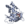
| Title | Cryo-EM Structure of OmpC3-MlaA-MlaC Complex in MSP2N2 Nanodiscs | ||||||
 Components Components |
| ||||||
 Keywords Keywords | LIPID TRANSPORT / bacteria / outer membrane / phospholipid / lipid asymmetry / membrane protein / protein complex structure / channel | ||||||
| Function / homology |  Function and homology information Function and homology informationintermembrane phospholipid transfer / phospholipid transport / porin activity / pore complex / cell outer membrane / outer membrane-bounded periplasmic space / virus receptor activity / monoatomic ion transmembrane transport / receptor-mediated virion attachment to host cell / DNA damage response ...intermembrane phospholipid transfer / phospholipid transport / porin activity / pore complex / cell outer membrane / outer membrane-bounded periplasmic space / virus receptor activity / monoatomic ion transmembrane transport / receptor-mediated virion attachment to host cell / DNA damage response / metal ion binding / identical protein binding Similarity search - Function | ||||||
| Biological species |  | ||||||
| Method | ELECTRON MICROSCOPY / single particle reconstruction / cryo EM / Resolution: 3.25 Å | ||||||
 Authors Authors | Yeow, J. / Luo, M. / Chng, S.S. | ||||||
| Funding support |  Singapore, 1items Singapore, 1items
| ||||||
 Citation Citation | Journal: Nat Commun / Year: 2023 Title: Molecular mechanism of phospholipid transport at the bacterial outer membrane interface. Authors: Jiang Yeow / Min Luo / Shu-Sin Chng / Abstract: The outer membrane (OM) of Gram-negative bacteria is an asymmetric lipid bilayer with outer leaflet lipopolysaccharides and inner leaflet phospholipids (PLs). This unique lipid asymmetry renders the ...The outer membrane (OM) of Gram-negative bacteria is an asymmetric lipid bilayer with outer leaflet lipopolysaccharides and inner leaflet phospholipids (PLs). This unique lipid asymmetry renders the OM impermeable to external insults, including antibiotics and bile salts. To maintain this barrier, the OmpC-Mla system removes mislocalized PLs from the OM outer leaflet, and transports them to the inner membrane (IM); in the first step, the OmpC-MlaA complex transfers PLs to the periplasmic chaperone MlaC, but mechanistic details are lacking. Here, we biochemically and structurally characterize the MlaA-MlaC transient complex. We map the interaction surfaces between MlaA and MlaC in Escherichia coli, and show that electrostatic interactions are important for MlaC recruitment to the OM. We further demonstrate that interactions with MlaC modulate conformational states in MlaA. Finally, we solve a 2.9-Å cryo-EM structure of a disulfide-trapped OmpC-MlaA-MlaC complex in nanodiscs, reinforcing the mechanism of MlaC recruitment, and highlighting membrane thinning as a plausible strategy for directing lipids for transport. Our work offers critical insights into retrograde PL transport by the OmpC-Mla system in maintaining OM lipid asymmetry. | ||||||
| History |
|
- Structure visualization
| Structure viewer | Molecule: MolmilJmol/JSmol |
|---|
- Downloads & links
-Download
| PDBx/mmCIF format | 8i8x.cif.gz | 294.3 KB | Display | PDBx/mmCIF format |
|---|---|---|---|---|
| PDB format | pdb8i8x.ent.gz | Display | PDB format | |
| PDBx/mmJSON format | 8i8x.json.gz | Tree view | PDBx/mmJSON format | |
| Others |  Other downloads Other downloads |
-Validation report
| Arichive directory | https://data.pdbj.org/pub/pdb/validation_reports/i8/8i8xftp://data.pdbj.org/pub/pdb/validation_reports/i8/8i8x | HTTPS FTP |
|---|
-Related structure data
| Related structure data |  35253MC  8i8rC C: citing same article ( M: map data used to model this data |
|---|---|
| Similar structure data |


-Links
 PDBj
PDBj


- Assembly
| Deposited unit | 
|
|---|---|
| 1 |
|
-Components
| #1: Protein | Mass: 38336.242 Da / Num. of mol.: 3 Source method: isolated from a genetically manipulated source Source: (gene. exp.) #2: Protein | | Mass: 26298.336 Da / Num. of mol.: 1 / Mutation: Q205C Source method: isolated from a genetically manipulated source Source: (gene. exp.) #3: Protein | | Mass: 22903.922 Da / Num. of mol.: 1 / Mutation: V171C Source method: isolated from a genetically manipulated source Source: (gene. exp.) #4: Chemical |   Mass: 2238.718 Da / Num. of mol.: 3 / Source method: obtained synthetically / Formula: C110H202N2O39P2 Mass: 2238.718 Da / Num. of mol.: 3 / Source method: obtained synthetically / Formula: C110H202N2O39P2Has ligand of interest | N | |
|---|
-Experimental details
-Experiment
| Experiment | Method: ELECTRON MICROSCOPY |
|---|---|
| EM experiment | Aggregation state: PARTICLE / 3D reconstruction method: single particle reconstruction |
- Sample preparation
| Component |
| ||||||||||||||||||||||||||||
|---|---|---|---|---|---|---|---|---|---|---|---|---|---|---|---|---|---|---|---|---|---|---|---|---|---|---|---|---|---|
| Molecular weight |
| ||||||||||||||||||||||||||||
| Source (natural) |
| ||||||||||||||||||||||||||||
| Source (recombinant) |
| ||||||||||||||||||||||||||||
| Buffer solution | pH: 8 Details: Tris-buffered saline (TBS) buffer (20 mM Tris HCl pH 8.0, 150 mM NaCl) | ||||||||||||||||||||||||||||
| Buffer component |
| ||||||||||||||||||||||||||||
| Specimen | Conc.: 12 mg/ml / Embedding applied: NO / Shadowing applied: NO / Staining applied: NO / Vitrification applied: YES | ||||||||||||||||||||||||||||
| Specimen support | Grid material: COPPER / Grid mesh size: 200 divisions/in. / Grid type: Quantifoil R1.2/1.3 | ||||||||||||||||||||||||||||
| Vitrification | Instrument: FEI VITROBOT MARK IV / Cryogen name: ETHANE / Humidity: 100 % / Chamber temperature: 277 K |
- Electron microscopy imaging
| Experimental equipment |  Model: Titan Krios / Image courtesy: FEI Company |
|---|---|
| Microscopy | Model: FEI TITAN KRIOS |
| Electron gun | Electron source:  FIELD EMISSION GUN / Accelerating voltage: 300 kV / Illumination mode: SPOT SCAN FIELD EMISSION GUN / Accelerating voltage: 300 kV / Illumination mode: SPOT SCAN |
| Electron lens | Mode: BRIGHT FIELD / Nominal magnification: 105000 X / Nominal defocus max: 2000 nm / Nominal defocus min: 800 nm / Cs: 2.7 mm |
| Specimen holder | Cryogen: NITROGEN / Specimen holder model: FEI TITAN KRIOS AUTOGRID HOLDER |
| Image recording | Average exposure time: 5.99 sec. / Electron dose: 90 e/Å2 / Film or detector model: GATAN K3 (6k x 4k) / Num. of grids imaged: 1 / Num. of real images: 6018 Details: Images were collected in movie-mode at 50 frames per image |
| EM imaging optics | Energyfilter name: GIF Tridiem 4K Details: Gatan GIF post-column energy filter operated in zero-loss mode Energyfilter slit width: 20 eV |
| Image scans | Sampling size: 5.2 µm / Width: 5760 / Height: 4092 |
- Processing
| EM software |
| ||||||||||||||||||||||||||||||||||||||||||||||||
|---|---|---|---|---|---|---|---|---|---|---|---|---|---|---|---|---|---|---|---|---|---|---|---|---|---|---|---|---|---|---|---|---|---|---|---|---|---|---|---|---|---|---|---|---|---|---|---|---|---|
| CTF correction | Details: Patch CTF routine in cryoSPARC / Type: PHASE FLIPPING AND AMPLITUDE CORRECTION | ||||||||||||||||||||||||||||||||||||||||||||||||
| Particle selection | Num. of particles selected: 2434319 Details: We performed automated particle picking using Blob and Template Picker for initial template picking and final sampling respectively. | ||||||||||||||||||||||||||||||||||||||||||||||||
| Symmetry | Point symmetry: C1 (asymmetric) | ||||||||||||||||||||||||||||||||||||||||||||||||
| 3D reconstruction | Resolution: 3.25 Å / Resolution method: FSC 0.143 CUT-OFF / Num. of particles: 76463 / Algorithm: BACK PROJECTION / Symmetry type: POINT | ||||||||||||||||||||||||||||||||||||||||||||||||
| Atomic model building | B value: 69.1 / Protocol: FLEXIBLE FIT / Space: REAL / Target criteria: Cross-correlation coefficient | ||||||||||||||||||||||||||||||||||||||||||||||||
| Atomic model building | 3D fitting-ID: 1 / Details: The initial model consisted of the chains A,B,C,D, and chain A for PDB entry 5UWA / Source name: PDB / Type: experimental model
| ||||||||||||||||||||||||||||||||||||||||||||||||
| Refine LS restraints |
|