 Movie
Movie Controller


+ Open data

- Basic information
| Entry | Database: PDB / ID: 8hqz | |||||||||||||||||||||||||||||||||||||||||||||||||||||||||||||||
|---|---|---|---|---|---|---|---|---|---|---|---|---|---|---|---|---|---|---|---|---|---|---|---|---|---|---|---|---|---|---|---|---|---|---|---|---|---|---|---|---|---|---|---|---|---|---|---|---|---|---|---|---|---|---|---|---|---|---|---|---|---|---|---|---|
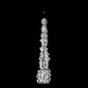
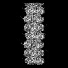
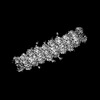
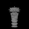
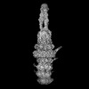
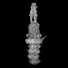
| Title | Baseplate of DT57C bacteriophage in the full state | |||||||||||||||||||||||||||||||||||||||||||||||||||||||||||||||
 Components Components |
| |||||||||||||||||||||||||||||||||||||||||||||||||||||||||||||||
 Keywords Keywords | VIRAL PROTEIN / Baseplate / T5 / VIRUS | |||||||||||||||||||||||||||||||||||||||||||||||||||||||||||||||
| Function / homology |  Function and homology information Function and homology information | |||||||||||||||||||||||||||||||||||||||||||||||||||||||||||||||
| Biological species |  Escherichia phage DT57C (virus) Escherichia phage DT57C (virus) | |||||||||||||||||||||||||||||||||||||||||||||||||||||||||||||||
| Method | ELECTRON MICROSCOPY / single particle reconstruction / cryo EM / Resolution: 3.8 Å | |||||||||||||||||||||||||||||||||||||||||||||||||||||||||||||||
 Authors Authors | Ayala, R. / Moiseenko, A.V. / Chen, T.H. / Kulikov, E.E. / Golomidova, A.K. / Orekhov, P.S. / Street, M.A. / Sokolova, O.S. / Letarov, A.V. / Wolf, M. | |||||||||||||||||||||||||||||||||||||||||||||||||||||||||||||||
| Funding support |  Russian Federation, Russian Federation,  Japan, 4items Japan, 4items
| |||||||||||||||||||||||||||||||||||||||||||||||||||||||||||||||
 Citation Citation | Journal: Nat Commun / Year: 2023 Title: Nearly complete structure of bacteriophage DT57C reveals architecture of head-to-tail interface and lateral tail fibers. Authors: Rafael Ayala / Andrey V Moiseenko / Ting-Hua Chen / Eugene E Kulikov / Alla K Golomidova / Philipp S Orekhov / Maya A Street / Olga S Sokolova / Andrey V Letarov / Matthias Wolf /   Abstract: The T5 family of viruses are tailed bacteriophages characterized by a long non-contractile tail. The bacteriophage DT57C is closely related to the paradigmal T5 phage, though it recognizes a ...The T5 family of viruses are tailed bacteriophages characterized by a long non-contractile tail. The bacteriophage DT57C is closely related to the paradigmal T5 phage, though it recognizes a different receptor (BtuB) and features highly divergent lateral tail fibers (LTF). Considerable portions of T5-like phages remain structurally uncharacterized. Here, we present the structure of DT57C determined by cryo-EM, and an atomic model of the virus, which was further explored using all-atom molecular dynamics simulations. The structure revealed a unique way of LTF attachment assisted by a dodecameric collar protein LtfC, and an unusual composition of the phage neck constructed of three protein rings. The tape measure protein (TMP) is organized within the tail tube in a three-stranded parallel α-helical coiled coil which makes direct contact with the genomic DNA. The presence of the C-terminal fragment of the TMP that remains within the tail tip suggests that the tail tip complex returns to its original state after DNA ejection. Our results provide a complete atomic structure of a T5-like phage, provide insights into the process of DNA ejection as well as a structural basis for the design of engineered phages and future mechanistic studies. | |||||||||||||||||||||||||||||||||||||||||||||||||||||||||||||||
| History |
|
- Structure visualization
| Structure viewer | Molecule: MolmilJmol/JSmol |
|---|
- Downloads & links
-Download
| PDBx/mmCIF format | 8hqz.cif.gz | 574.5 KB | Display | PDBx/mmCIF format |
|---|---|---|---|---|
| PDB format | pdb8hqz.ent.gz | Display | PDB format | |
| PDBx/mmJSON format | 8hqz.json.gz | Tree view | PDBx/mmJSON format | |
| Others |  Other downloads Other downloads |
-Validation report
| Summary document | 8hqz_validation.pdf.gz | 1.3 MB | Display | wwPDB validaton report |
|---|---|---|---|---|
| Full document | 8hqz_full_validation.pdf.gz | 1.3 MB | Display | |
| Data in XML | 8hqz_validation.xml.gz | 81.8 KB | Display | |
| Data in CIF | 8hqz_validation.cif.gz | 130.3 KB | Display | |
| Arichive directory | https://data.pdbj.org/pub/pdb/validation_reports/hq/8hqzftp://data.pdbj.org/pub/pdb/validation_reports/hq/8hqz | HTTPS FTP |
-Related structure data
| Related structure data | 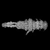 34955MC  8ho3C  8hqkC  8hqoC  8hreC  8hrgC C: citing same article ( M: map data used to model this data |
|---|---|
| Similar structure data |


-Links
 PDBj
PDBj- Assembly
| Deposited unit | 
|
|---|---|
| 1 | 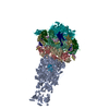
|
| 2 |
|
| 3 | 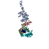
|
| Symmetry | Point symmetry: (Schoenflies symbol: C3 (3 fold cyclic)) |
-Components
-Protein , 5 types, 6 molecules AHILkp
| #1: Protein | Mass: 107114.055 Da / Num. of mol.: 1 / Source method: isolated from a natural source / Source: (natural) Escherichia phage DT57C (virus) / References: UniProt: A0A0A7RSL1 | ||||||
|---|---|---|---|---|---|---|---|
| #2: Protein | Mass: 22666.484 Da / Num. of mol.: 2 / Source method: isolated from a natural source / Source: (natural) Escherichia phage DT57C (virus) / References: UniProt: A0A0A7RSH9#3: Protein | | Mass: 34513.672 Da / Num. of mol.: 1 / Source method: isolated from a natural source / Source: (natural) Escherichia phage DT57C (virus) / References: UniProt: A0A0A7RSL6#6: Protein | | Mass: 132626.406 Da / Num. of mol.: 1 / Source method: isolated from a natural source / Source: (natural) Escherichia phage DT57C (virus) / References: UniProt: A0A0A7RZ92#7: Protein | | Mass: 50759.051 Da / Num. of mol.: 1 / Source method: isolated from a natural source / Source: (natural) Escherichia phage DT57C (virus) / References: UniProt: A0A0A7RSI5 |
-L-shaped tail fiber ... , 2 types, 7 molecules QRSTdef
| #4: Protein | Mass: 15384.226 Da / Num. of mol.: 4 / Source method: isolated from a natural source / Source: (natural) Escherichia phage DT57C (virus) / References: UniProt: A0A0A7RUJ8#5: Protein | Mass: 113712.461 Da / Num. of mol.: 3 / Source method: isolated from a natural source / Source: (natural) Escherichia phage DT57C (virus) / References: UniProt: A0A0A7RZ88 |
|---|
-Details
| Has protein modification | Y |
|---|
-Experimental details
-Experiment
| Experiment | Method: ELECTRON MICROSCOPY |
|---|---|
| EM experiment | Aggregation state: PARTICLE / 3D reconstruction method: single particle reconstruction |
- Sample preparation
| Component | Name: Escherichia phage DT57C / Type: VIRUS / Entity ID: all / Source: NATURAL |
|---|---|
| Source (natural) | Organism: Escherichia phage DT57C (virus) |
| Details of virus | Empty: NO / Enveloped: NO / Isolate: STRAIN / Type: VIRION |
| Buffer solution | pH: 7.5 |
| Specimen | Embedding applied: NO / Shadowing applied: NO / Staining applied: NO / Vitrification applied: YES |
| Vitrification | Cryogen name: ETHANE-PROPANE |
- Electron microscopy imaging
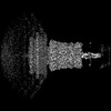
| Experimental equipment |  Model: Titan Krios / Image courtesy: FEI Company |
|---|---|
| Microscopy | Model: FEI TITAN KRIOS |
| Electron gun | Electron source:  FIELD EMISSION GUN / Accelerating voltage: 300 kV / Illumination mode: FLOOD BEAM FIELD EMISSION GUN / Accelerating voltage: 300 kV / Illumination mode: FLOOD BEAM |
| Electron lens | Mode: BRIGHT FIELD / Nominal defocus max: 4000 nm / Nominal defocus min: 1000 nm |
| Image recording | Electron dose: 67 e/Å2 / Film or detector model: FEI FALCON III (4k x 4k) |
- Processing
| EM software |
| ||||||||||||||||||||||||
|---|---|---|---|---|---|---|---|---|---|---|---|---|---|---|---|---|---|---|---|---|---|---|---|---|---|
| CTF correction | Type: PHASE FLIPPING AND AMPLITUDE CORRECTION | ||||||||||||||||||||||||
| 3D reconstruction | Resolution: 3.8 Å / Resolution method: FSC 0.143 CUT-OFF / Num. of particles: 42697 / Symmetry type: POINT | ||||||||||||||||||||||||
| Refine LS restraints |
|
