 Movie
Movie Controller

[English] 日本語
 Yorodumi
- PDB-8fsj: Cryo-EM structure of engineered hepatitis C virus E1E2 ectodomain... -
+ Open data

- Basic information
| Entry | Database: PDB / ID: 8fsj | ||||||
|---|---|---|---|---|---|---|---|
| Title | Cryo-EM structure of engineered hepatitis C virus E1E2 ectodomain in complex with antibodies AR4A, HEPC74, and IGH520 | ||||||
 Components Components |
| ||||||
 Keywords Keywords | VIRAL PROTEIN / HCV / E1E2 | ||||||
| Function / homology |  Function and homology information Function and homology informationhost cell mitochondrial membrane / host cell lipid droplet / viral nucleocapsid / clathrin-dependent endocytosis of virus by host cell / symbiont-mediated suppression of host innate immune response / host cell endoplasmic reticulum membrane / ribonucleoprotein complex / fusion of virus membrane with host endosome membrane / viral envelope / virion attachment to host cell ...host cell mitochondrial membrane / host cell lipid droplet / viral nucleocapsid / clathrin-dependent endocytosis of virus by host cell / symbiont-mediated suppression of host innate immune response / host cell endoplasmic reticulum membrane / ribonucleoprotein complex / fusion of virus membrane with host endosome membrane / viral envelope / virion attachment to host cell / host cell nucleus / virion membrane / structural molecule activity / RNA binding Similarity search - Function | ||||||
| Biological species |  Hepacivirus C Hepacivirus Csynthetic construct (others)  Homo sapiens (human) Homo sapiens (human) | ||||||
| Method | ELECTRON MICROSCOPY / single particle reconstruction / cryo EM / Resolution: 3.65 Å | ||||||
 Authors Authors | Metcalf, M.C. / Ofek, G. | ||||||
| Funding support |  United States, 1items United States, 1items
| ||||||
 Citation Citation | Journal: Nat Commun / Year: 2023 Title: Structure of engineered hepatitis C virus E1E2 ectodomain in complex with neutralizing antibodies. Authors: Matthew C Metcalf / Benjamin M Janus / Rui Yin / Ruixue Wang / Johnathan D Guest / Edwin Pozharski / Mansun Law / Roy A Mariuzza / Eric A Toth / Brian G Pierce / Thomas R Fuerst / Gilad Ofek / Abstract: Hepatitis C virus (HCV) is a major global health burden as the leading causative agent of chronic liver disease and hepatocellular carcinoma. While the main antigenic target for HCV-neutralizing ...Hepatitis C virus (HCV) is a major global health burden as the leading causative agent of chronic liver disease and hepatocellular carcinoma. While the main antigenic target for HCV-neutralizing antibodies is the membrane-associated E1E2 surface glycoprotein, the development of effective vaccines has been hindered by complications in the biochemical preparation of soluble E1E2 ectodomains. Here, we present a cryo-EM structure of an engineered, secreted E1E2 ectodomain of genotype 1b in complex with neutralizing antibodies AR4A, HEPC74, and IGH520. Structural characterization of the E1 subunit and C-terminal regions of E2 reveal an overall architecture of E1E2 that concurs with that observed for non-engineered full-length E1E2. Analysis of the AR4A epitope within a region of E2 that bridges between the E2 core and E1 defines the structural basis for its broad neutralization. Our study presents the structure of an E1E2 complex liberated from membrane via a designed scaffold, one that maintains all essential structural features of native E1E2. The study advances the understanding of the E1E2 heterodimer structure, crucial for the rational design of secreted E1E2 antigens in vaccine development. | ||||||
| History |
|
- Structure visualization
| Structure viewer | Molecule: MolmilJmol/JSmol |
|---|
- Downloads & links
-Download
| PDBx/mmCIF format | 8fsj.cif.gz | 243.6 KB | Display | PDBx/mmCIF format |
|---|---|---|---|---|
| PDB format | pdb8fsj.ent.gz | 187.8 KB | Display | PDB format |
| PDBx/mmJSON format | 8fsj.json.gz | Tree view | PDBx/mmJSON format | |
| Others |  Other downloads Other downloads |
-Validation report
| Arichive directory | https://data.pdbj.org/pub/pdb/validation_reports/fs/8fsjftp://data.pdbj.org/pub/pdb/validation_reports/fs/8fsj | HTTPS FTP |
|---|
-Related structure data
| Related structure data | 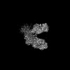 29419MC M: map data used to model this data C: citing same article ( |
|---|---|
| Similar structure data |
-Links
 PDBj
PDBj


- Assembly
| Deposited unit | 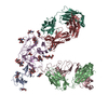
|
|---|---|
| 1 |
|
-Components
-Protein , 3 types, 3 molecules EAC
| #1: Protein | Mass: 42975.371 Da / Num. of mol.: 1 Source method: isolated from a genetically manipulated source Source: (gene. exp.) Hepacivirus C, (gene. exp.) synthetic construct (others)Strain: 1b09 / Plasmid: pCMV / Cell line (production host): HEK293 / Production host: Homo sapiens (human) / Tissue (production host): kidney / References: UniProt: A0A2P0NE15 |
|---|---|
| #4: Protein | Mass: 22951.379 Da / Num. of mol.: 1 / Fragment: residues 22-179 Source method: isolated from a genetically manipulated source Source: (gene. exp.) Hepacivirus C, (gene. exp.) synthetic construct (others)Strain: 1b09 / Plasmid: pCMV / Cell line (production host): HEK293 / Production host: Homo sapiens (human) / Tissue (production host): kidney / References: UniProt: A0A2P0NE34 |
| #6: Protein | Mass: 31862.697 Da / Num. of mol.: 1 Source method: isolated from a genetically manipulated source Source: (gene. exp.) Homo sapiens (human) / Plasmid: pCMV / Cell line (production host): HEK293 / Production host: Homo sapiens (human) / Tissue (production host): kidney |
-Antibody , 3 types, 3 molecules HLB
| #2: Antibody | Mass: 27644.807 Da / Num. of mol.: 1 Source method: isolated from a genetically manipulated source Source: (gene. exp.) Homo sapiens (human) / Plasmid: pCMV / Cell line (production host): HEK293 / Production host: Homo sapiens (human) / Tissue (production host): kidney |
|---|---|
| #3: Antibody | Mass: 23470.111 Da / Num. of mol.: 1 Source method: isolated from a genetically manipulated source Source: (gene. exp.) Homo sapiens (human) / Plasmid: pCMV / Cell line (production host): HEK293 / Production host: Homo sapiens (human) / Tissue (production host): kidney |
| #5: Antibody | Mass: 25743.781 Da / Num. of mol.: 1 Source method: isolated from a genetically manipulated source Source: (gene. exp.) Homo sapiens (human) / Plasmid: pCMV / Cell line (production host): HEK293 / Production host: Homo sapiens (human) / Tissue (production host): kidney |
-Sugars , 3 types, 14 molecules 
| #7: Polysaccharide | Source method: isolated from a genetically manipulated source #8: Polysaccharide | beta-D-mannopyranose-(1-4)-2-acetamido-2-deoxy-beta-D-glucopyranose-(1-4)-2-acetamido-2-deoxy-beta- ...beta-D-mannopyranose-(1-4)-2-acetamido-2-deoxy-beta-D-glucopyranose-(1-4)-2-acetamido-2-deoxy-beta-D-glucopyranose Source method: isolated from a genetically manipulated source #9: Sugar | ChemComp-NAG /  Type: D-saccharide, beta linking / Mass: 221.208 Da / Num. of mol.: 6 / Source method: obtained synthetically / Formula: C8H15NO6 / Feature type: SUBJECT OF INVESTIGATION Type: D-saccharide, beta linking / Mass: 221.208 Da / Num. of mol.: 6 / Source method: obtained synthetically / Formula: C8H15NO6 / Feature type: SUBJECT OF INVESTIGATION |
|---|
-Details
| Has ligand of interest | Y |
|---|---|
| Has protein modification | Y |
-Experimental details
-Experiment
| Experiment | Method: ELECTRON MICROSCOPY |
|---|---|
| EM experiment | Aggregation state: PARTICLE / 3D reconstruction method: single particle reconstruction |
- Sample preparation
| Component | Name: HCV E1E2 ectodomain in complex with AR4A, HEPC74, and IGH520 Type: COMPLEX / Entity ID: #1-#6 / Source: RECOMBINANT |
|---|---|
| Molecular weight | Value: 0.250 MDa / Experimental value: NO |
| Source (natural) | Organism: Hepacivirus C |
| Source (recombinant) | Organism: Homo sapiens (human) / Cell: HEK293 |
| Buffer solution | pH: 7.4 |
| Specimen | Conc.: 1 mg/ml / Embedding applied: NO / Shadowing applied: NO / Staining applied: NO / Vitrification applied: YES |
| Specimen support | Grid material: GOLD / Grid mesh size: 400 divisions/in. / Grid type: Quantifoil R1.2/1.3 |
| Vitrification | Cryogen name: ETHANE / Humidity: 100 % |
- Electron microscopy imaging
| Microscopy | Model: TFS GLACIOS |
|---|---|
| Electron gun | Electron source:  FIELD EMISSION GUN / Accelerating voltage: 200 kV / Illumination mode: FLOOD BEAM FIELD EMISSION GUN / Accelerating voltage: 200 kV / Illumination mode: FLOOD BEAM |
| Electron lens | Mode: BRIGHT FIELD / Nominal defocus max: 2500 nm / Nominal defocus min: 500 nm |
| Image recording | Electron dose: 47 e/Å2 / Film or detector model: GATAN K3 (6k x 4k) |
- Processing
| Software | Name: PHENIX / Version: 1.19.2_4158: / Classification: refinement | ||||||||||||||||||||||||
|---|---|---|---|---|---|---|---|---|---|---|---|---|---|---|---|---|---|---|---|---|---|---|---|---|---|
| EM software |
| ||||||||||||||||||||||||
| CTF correction | Type: PHASE FLIPPING ONLY | ||||||||||||||||||||||||
| Particle selection | Num. of particles selected: 2620845 | ||||||||||||||||||||||||
| 3D reconstruction | Resolution: 3.65 Å / Resolution method: FSC 0.143 CUT-OFF / Num. of particles: 143576 / Symmetry type: POINT | ||||||||||||||||||||||||
| Refine LS restraints |
|
