 Movie
Movie Controller

[English] 日本語
 Yorodumi
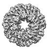
- PDB-8emh: CryoEM characterization of a unique AAA+ BrxL phage restriction factor -
+ Open data

- Basic information
| Entry | Database: PDB / ID: 8emh | ||||||
|---|---|---|---|---|---|---|---|
| Title | CryoEM characterization of a unique AAA+ BrxL phage restriction factor | ||||||
 Components Components |
| ||||||
 Keywords Keywords | ANTIMICROBIAL PROTEIN / phage restriction factor / AAA+ protein | ||||||
| Function / homology |  Function and homology information Function and homology informationATP-dependent peptidase activity / protein catabolic process / serine-type endopeptidase activity / proteolysis / ATP binding Similarity search - Function | ||||||
| Biological species |  Acinetobacter sp. NEB 394 (bacteria) Acinetobacter sp. NEB 394 (bacteria)synthetic construct (others) | ||||||
| Method | ELECTRON MICROSCOPY / single particle reconstruction / cryo EM / Resolution: 3.63 Å | ||||||
 Authors Authors | Shen, B.W. / Stoddard, B.L. | ||||||
| Funding support |  United States, 1items United States, 1items
| ||||||
 Citation Citation | Journal: Nucleic Acids Res / Year: 2023 Title: Structure, substrate binding and activity of a unique AAA+ protein: the BrxL phage restriction factor. Authors: Betty W Shen / Lindsey A Doyle / Rachel Werther / Abigail A Westburg / Daniel P Bies / Stephanie I Walter / Yvette A Luyten / Richard D Morgan / Barry L Stoddard / Brett K Kaiser / Abstract: Bacteriophage exclusion ('BREX') systems are multi-protein complexes encoded by a variety of bacteria and archaea that restrict phage by an unknown mechanism. One BREX factor, termed BrxL, has been ...Bacteriophage exclusion ('BREX') systems are multi-protein complexes encoded by a variety of bacteria and archaea that restrict phage by an unknown mechanism. One BREX factor, termed BrxL, has been noted to display sequence similarity to various AAA+ protein factors including Lon protease. In this study we describe multiple CryoEM structures of BrxL that demonstrate it to be a chambered, ATP-dependent DNA binding protein. The largest BrxL assemblage corresponds to a dimer of heptamers in the absence of bound DNA, versus a dimer of hexamers when DNA is bound in its central pore. The protein displays DNA-dependent ATPase activity, and ATP binding promotes assembly of the complex on DNA. Point mutations within several regions of the protein-DNA complex alter one or more in vitro behaviors and activities, including ATPase activity and ATP-dependent association with DNA. However, only the disruption of the ATPase active site fully eliminates phage restriction, indicating that other mutations can still complement BrxL function within the context of an otherwise intact BREX system. BrxL displays significant structural homology to MCM subunits (the replicative helicase in archaea and eukaryotes), implying that it and other BREX factors may collaborate to disrupt initiation of phage DNA replication. | ||||||
| History |
|
- Structure visualization
| Structure viewer | Molecule: MolmilJmol/JSmol |
|---|
- Downloads & links
-Download
| PDBx/mmCIF format | 8emh.cif.gz | 1.3 MB | Display | PDBx/mmCIF format |
|---|---|---|---|---|
| PDB format | pdb8emh.ent.gz | 1.1 MB | Display | PDB format |
| PDBx/mmJSON format | 8emh.json.gz | Tree view | PDBx/mmJSON format | |
| Others |  Other downloads Other downloads |
-Validation report
| Summary document | 8emh_validation.pdf.gz | 1.5 MB | Display | wwPDB validaton report |
|---|---|---|---|---|
| Full document | 8emh_full_validation.pdf.gz | 1.6 MB | Display | |
| Data in XML | 8emh_validation.xml.gz | 213.2 KB | Display | |
| Data in CIF | 8emh_validation.cif.gz | 319 KB | Display | |
| Arichive directory | https://data.pdbj.org/pub/pdb/validation_reports/em/8emhftp://data.pdbj.org/pub/pdb/validation_reports/em/8emh | HTTPS FTP |
-Related structure data
| Related structure data | 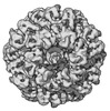 28248MC 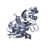 8eilC  8emcC C: citing same article ( M: map data used to model this data |
|---|---|
| Similar structure data | |
| Experimental dataset #1 | Data reference: 10.2210/pdb8emc/pdb. |
-Links
 PDBj
PDBj


- Assembly
| Deposited unit | 
|
|---|---|
| 1 |
|
-Components
| #1: Protein | Mass: 75702.555 Da / Num. of mol.: 12 Source method: isolated from a genetically manipulated source Source: (gene. exp.) Acinetobacter sp. NEB 394 (bacteria) / Gene: brxL, HUK62_18280 / Production host: #2: DNA chain | | Mass: 19689.639 Da / Num. of mol.: 1 / Source method: obtained synthetically / Source: (synth.) synthetic construct (others) #3: DNA chain | | Mass: 19485.461 Da / Num. of mol.: 1 / Source method: obtained synthetically / Source: (synth.) synthetic construct (others) |
|---|
-Experimental details
-Experiment
| Experiment | Method: ELECTRON MICROSCOPY |
|---|---|
| EM experiment | Aggregation state: PARTICLE / 3D reconstruction method: single particle reconstruction |
- Sample preparation
| Component | Name: complex of walkerB mutant of BrxL with random sequence of DNA fragments Type: COMPLEX Details: 12 strands of KrxL E280Q mutant bound to short random sequence DNA fragments. Entity ID: all / Source: RECOMBINANT |
|---|---|
| Molecular weight | Value: 0.950 MDa / Experimental value: NO |
| Source (natural) | Organism: Acinetobacter (bacteria) |
| Source (recombinant) | Organism: |
| Buffer solution | pH: 8 / Details: 20 mM TrisHCl, 150 mM NaCl |
| Specimen | Embedding applied: NO / Shadowing applied: NO / Staining applied: NO / Vitrification applied: YES / Details: The sample was mono-disperse |
| Specimen support | Details: 40 / Grid material: COPPER / Grid mesh size: 400 divisions/in. / Grid type: Quantifoil R1.2/1.3 |
| Vitrification | Cryogen name: ETHANE / Humidity: 95 % / Chamber temperature: 275 K |
- Electron microscopy imaging
| Experimental equipment |  Model: Talos Arctica / Image courtesy: FEI Company |
|---|---|
| Microscopy | Model: FEI TALOS ARCTICA |
| Electron gun | Electron source:  FIELD EMISSION GUN / Accelerating voltage: 200 kV / Illumination mode: FLOOD BEAM FIELD EMISSION GUN / Accelerating voltage: 200 kV / Illumination mode: FLOOD BEAM |
| Electron lens | Mode: BRIGHT FIELD / Nominal defocus max: 5000 nm / Nominal defocus min: 1200 nm / Calibrated defocus min: 12100 nm / Cs: 2.7 mm / C2 aperture diameter: 70 µm / Alignment procedure: COMA FREE |
| Specimen holder | Cryogen: NITROGEN / Specimen holder model: FEI TITAN KRIOS AUTOGRID HOLDER / Temperature (max): 70 K |
| Image recording | Average exposure time: 2 sec. / Electron dose: 30 e/Å2 / Detector mode: SUPER-RESOLUTION / Film or detector model: DIRECT ELECTRON DE-10 (5k x 4k) / Num. of grids imaged: 2 |
| Image scans | Movie frames/image: 50 / Used frames/image: 1-49 |
- Processing
| Software | Name: PHENIX / Version: 1.19.2_4158: / Classification: refinement | ||||||||||||||||||||||||
|---|---|---|---|---|---|---|---|---|---|---|---|---|---|---|---|---|---|---|---|---|---|---|---|---|---|
| EM software |
| ||||||||||||||||||||||||
| CTF correction | Type: PHASE FLIPPING AND AMPLITUDE CORRECTION | ||||||||||||||||||||||||
| Particle selection | Num. of particles selected: 3241183 | ||||||||||||||||||||||||
| Symmetry | Point symmetry: C1 (asymmetric) | ||||||||||||||||||||||||
| 3D reconstruction | Resolution: 3.63 Å / Resolution method: FSC 0.143 CUT-OFF / Num. of particles: 229430 / Num. of class averages: 4 / Symmetry type: POINT | ||||||||||||||||||||||||
| Refine LS restraints |
|
