Movie
Movie Controller

[English] 日本語
 Yorodumi
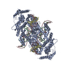
- PDB-8edg: Cryo-EM structure of the Hermes transposase bound to two left-end... -
+ Open data

- Basic information
| Entry | Database: PDB / ID: 8edg | ||||||
|---|---|---|---|---|---|---|---|
| Title | Cryo-EM structure of the Hermes transposase bound to two left-ends of its DNA transposon | ||||||
 Components Components |
| ||||||
 Keywords Keywords | RECOMBINATION/DNA / transposase / transpososome / BED domain / protein-DNA complex / RECOMBINATION / RECOMBINATION-DNA complex | ||||||
| Function / homology |  Function and homology information Function and homology informationprotein dimerization activity / regulation of transcription by RNA polymerase II / DNA binding / zinc ion binding / nucleus Similarity search - Function | ||||||
| Biological species |  Musca domestica (house fly) Musca domestica (house fly) | ||||||
| Method | ELECTRON MICROSCOPY / single particle reconstruction / cryo EM / Resolution: 4.64 Å | ||||||
 Authors Authors | Lannes, L. / Dyda, F. | ||||||
| Funding support |  United States, 1items United States, 1items
| ||||||
 Citation Citation | Journal: Nat Commun / Year: 2023 Title: Zinc-finger BED domains drive the formation of the active Hermes transpososome by asymmetric DNA binding. Authors: Laurie Lannes / Christopher M Furman / Alison B Hickman / Fred Dyda / Abstract: The Hermes DNA transposon is a member of the eukaryotic hAT superfamily, and its transposase forms a ring-shaped tetramer of dimers. Our investigation, combining biochemical, crystallography and cryo- ...The Hermes DNA transposon is a member of the eukaryotic hAT superfamily, and its transposase forms a ring-shaped tetramer of dimers. Our investigation, combining biochemical, crystallography and cryo-electron microscopy, and in-cell assays, shows that the full-length Hermes octamer extensively interacts with its transposon left-end through multiple BED domains of three Hermes protomers contributed by three dimers explaining the role of the unusual higher-order assembly. By contrast, the right-end is bound to no BED domains at all. Thus, this work supports a model in which Hermes multimerizes to gather enough BED domains to find its left-end among the abundant genomic DNA, facilitating the subsequent interaction with the right-end. | ||||||
| History |
|
- Structure visualization
| Structure viewer | Molecule: MolmilJmol/JSmol |
|---|
- Downloads & links
-Download
| PDBx/mmCIF format | 8edg.cif.gz | 531.3 KB | Display | PDBx/mmCIF format |
|---|---|---|---|---|
| PDB format | pdb8edg.ent.gz | 422.8 KB | Display | PDB format |
| PDBx/mmJSON format | 8edg.json.gz | Tree view | PDBx/mmJSON format | |
| Others |  Other downloads Other downloads |
-Validation report
| Arichive directory | https://data.pdbj.org/pub/pdb/validation_reports/ed/8edgftp://data.pdbj.org/pub/pdb/validation_reports/ed/8edg | HTTPS FTP |
|---|
-Related structure data
| Related structure data |  28034MC  8eb5C 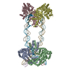 8sjdC M: map data used to model this data C: citing same article ( |
|---|---|
| Similar structure data |

-Links
 PDBj
PDBj


- Assembly
| Deposited unit | 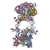
|
|---|---|
| 1 |
|
-Components
| #1: DNA chain | Mass: 2162.448 Da / Num. of mol.: 2 / Source method: obtained synthetically / Source: (synth.) Musca domestica (house fly)#2: DNA chain | Mass: 14173.139 Da / Num. of mol.: 2 / Source method: obtained synthetically / Source: (synth.) Musca domestica (house fly)#3: DNA chain | Mass: 16931.867 Da / Num. of mol.: 2 / Source method: obtained synthetically / Source: (synth.) Musca domestica (house fly)#4: Protein | Mass: 70210.570 Da / Num. of mol.: 6 / Mutation: Q2E,K128G Source method: isolated from a genetically manipulated source Source: (gene. exp.) Musca domestica (house fly) / Plasmid: pBAD/Myc-His / Production host:  #5: Chemical | ChemComp-ZN /   Mass: 65.409 Da / Num. of mol.: 6 / Source method: obtained synthetically / Formula: Zn Mass: 65.409 Da / Num. of mol.: 6 / Source method: obtained synthetically / Formula: ZnHas ligand of interest | Y | |
|---|
-Experimental details
-Experiment
| Experiment | Method: ELECTRON MICROSCOPY |
|---|---|
| EM experiment | Aggregation state: PARTICLE / 3D reconstruction method: single particle reconstruction |
- Sample preparation
| Component | Name: Two left-end Hermes transpososome / Type: COMPLEX Details: Hermes transposase tetramer of dimers complex bound to two transposon left-end DNAs. The complex was obtained by mixing the purified protein and the DNA. Entity ID: #1-#4 / Source: MULTIPLE SOURCES | ||||||||||||||||||||
|---|---|---|---|---|---|---|---|---|---|---|---|---|---|---|---|---|---|---|---|---|---|
| Molecular weight | Value: 0.627 MDa / Experimental value: NO | ||||||||||||||||||||
| Source (natural) | Organism: Musca domestica (house fly) | ||||||||||||||||||||
| Source (recombinant) | Organism: | ||||||||||||||||||||
| Buffer solution | pH: 7.5 | ||||||||||||||||||||
| Buffer component |
| ||||||||||||||||||||
| Specimen | Conc.: 0.2 mg/ml / Embedding applied: NO / Shadowing applied: NO / Staining applied: NO / Vitrification applied: YES Details: The complex was formed in vitro by mixing the purified protein with the DNA and further purified by size exclusion chromatography. | ||||||||||||||||||||
| Specimen support | Grid mesh size: 300 divisions/in. / Grid type: Quantifoil R1.2/1.3 | ||||||||||||||||||||
| Vitrification | Instrument: FEI VITROBOT MARK IV / Cryogen name: ETHANE / Humidity: 100 % / Chamber temperature: 298 K |
- Electron microscopy imaging
| Microscopy | Model: TFS GLACIOS |
|---|---|
| Electron gun | Electron source:  FIELD EMISSION GUN / Accelerating voltage: 200 kV / Illumination mode: FLOOD BEAM FIELD EMISSION GUN / Accelerating voltage: 200 kV / Illumination mode: FLOOD BEAM |
| Electron lens | Mode: BRIGHT FIELD / Nominal magnification: 130000 X / Nominal defocus max: 2500 nm / Nominal defocus min: 1000 nm |
| Specimen holder | Cryogen: NITROGEN |
| Image recording | Average exposure time: 2 sec. / Electron dose: 22.3 e/Å2 / Film or detector model: GATAN K3 (6k x 4k) / Num. of grids imaged: 2 / Num. of real images: 9500 |
- Processing
| Software | Name: PHENIX / Version: 1.19.2_4158: / Classification: refinement | ||||||||||||||||||||||||||||||||||||||||||||
|---|---|---|---|---|---|---|---|---|---|---|---|---|---|---|---|---|---|---|---|---|---|---|---|---|---|---|---|---|---|---|---|---|---|---|---|---|---|---|---|---|---|---|---|---|---|
| EM software |
| ||||||||||||||||||||||||||||||||||||||||||||
| CTF correction | Type: PHASE FLIPPING AND AMPLITUDE CORRECTION | ||||||||||||||||||||||||||||||||||||||||||||
| Particle selection | Num. of particles selected: 3850000 | ||||||||||||||||||||||||||||||||||||||||||||
| Symmetry | Point symmetry: C1 (asymmetric) | ||||||||||||||||||||||||||||||||||||||||||||
| 3D reconstruction | Resolution: 4.64 Å / Resolution method: FSC 0.143 CUT-OFF / Num. of particles: 143400 Details: The 3D reconstruction is not representative of the complex in solution. The Hermes transposase forms a tetramer of dimers assembled in a ring like shape. Due to their low resolution two ...Details: The 3D reconstruction is not representative of the complex in solution. The Hermes transposase forms a tetramer of dimers assembled in a ring like shape. Due to their low resolution two Hermes dimers have been partially or completely masked out during the processing. Only the opposite two Hermes dimers interacting with the DNAs have been fully reconstructed, along with the two DNAs and the two BED domains of a third Hermes dimer. Symmetry type: POINT | ||||||||||||||||||||||||||||||||||||||||||||
| Atomic model building | Protocol: FLEXIBLE FIT / Space: REAL | ||||||||||||||||||||||||||||||||||||||||||||
| Atomic model building |
| ||||||||||||||||||||||||||||||||||||||||||||
| Refinement | Cross valid method: NONE Stereochemistry target values: GeoStd + Monomer Library + CDL v1.2 | ||||||||||||||||||||||||||||||||||||||||||||
| Displacement parameters | Biso mean: 173.07 Å2 | ||||||||||||||||||||||||||||||||||||||||||||
| Refine LS restraints |
|