Movie
Movie Controller

[English] 日本語
 Yorodumi
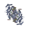
- PDB-8sjd: Cryo-EM structure of the Hermes transposase bound to two right-en... -
+ Open data

- Basic information
| Entry | Database: PDB / ID: 8sjd | ||||||
|---|---|---|---|---|---|---|---|
| Title | Cryo-EM structure of the Hermes transposase bound to two right-ends of its DNA transposon. | ||||||
 Components Components |
| ||||||
 Keywords Keywords | RECOMBINATION/DNA / transposase / transpososome / BED domain / protein-DNA complex / RECOMBINATION-DNA complex | ||||||
| Function / homology |  Function and homology information Function and homology informationprotein dimerization activity / regulation of transcription by RNA polymerase II / DNA binding / zinc ion binding / nucleus Similarity search - Function | ||||||
| Biological species |  Musca domestica (house fly) Musca domestica (house fly) | ||||||
| Method | ELECTRON MICROSCOPY / single particle reconstruction / cryo EM / Resolution: 5.1 Å | ||||||
 Authors Authors | Lannes, L. / Dyda, F. | ||||||
| Funding support |  United States, 1items United States, 1items
| ||||||
 Citation Citation | Journal: Nat Commun / Year: 2023 Title: Zinc-finger BED domains drive the formation of the active Hermes transpososome by asymmetric DNA binding. Authors: Laurie Lannes / Christopher M Furman / Alison B Hickman / Fred Dyda / Abstract: The Hermes DNA transposon is a member of the eukaryotic hAT superfamily, and its transposase forms a ring-shaped tetramer of dimers. Our investigation, combining biochemical, crystallography and cryo- ...The Hermes DNA transposon is a member of the eukaryotic hAT superfamily, and its transposase forms a ring-shaped tetramer of dimers. Our investigation, combining biochemical, crystallography and cryo-electron microscopy, and in-cell assays, shows that the full-length Hermes octamer extensively interacts with its transposon left-end through multiple BED domains of three Hermes protomers contributed by three dimers explaining the role of the unusual higher-order assembly. By contrast, the right-end is bound to no BED domains at all. Thus, this work supports a model in which Hermes multimerizes to gather enough BED domains to find its left-end among the abundant genomic DNA, facilitating the subsequent interaction with the right-end. | ||||||
| History |
|
- Structure visualization
| Structure viewer | Molecule: MolmilJmol/JSmol |
|---|
- Downloads & links
-Download
| PDBx/mmCIF format | 8sjd.cif.gz | 446.9 KB | Display | PDBx/mmCIF format |
|---|---|---|---|---|
| PDB format | pdb8sjd.ent.gz | 357.8 KB | Display | PDB format |
| PDBx/mmJSON format | 8sjd.json.gz | Tree view | PDBx/mmJSON format | |
| Others |  Other downloads Other downloads |
-Validation report
| Arichive directory | https://data.pdbj.org/pub/pdb/validation_reports/sj/8sjdftp://data.pdbj.org/pub/pdb/validation_reports/sj/8sjd | HTTPS FTP |
|---|
-Related structure data
| Related structure data |  40553MC  8eb5C 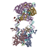 8edgC M: map data used to model this data C: citing same article ( |
|---|---|
| Similar structure data |

-Links
 PDBj
PDBj


- Assembly
| Deposited unit | 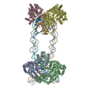
|
|---|---|
| 1 |
|
-Components
| #1: Protein | Mass: 70210.570 Da / Num. of mol.: 4 / Mutation: Q2E,K128G Source method: isolated from a genetically manipulated source Source: (gene. exp.) Musca domestica (house fly) / Plasmid: pBAD/Myc-His / Details (production host): no fusion tag / Production host:  #2: DNA chain | Mass: 16909.779 Da / Num. of mol.: 2 / Source method: obtained synthetically / Source: (synth.) Musca domestica (house fly)#3: DNA chain | Mass: 14197.199 Da / Num. of mol.: 2 / Source method: obtained synthetically / Source: (synth.) Musca domestica (house fly)#4: DNA chain | Mass: 2162.448 Da / Num. of mol.: 2 / Source method: obtained synthetically / Source: (synth.) Musca domestica (house fly) |
|---|
-Experimental details
-Experiment
| Experiment | Method: ELECTRON MICROSCOPY |
|---|---|
| EM experiment | Aggregation state: PARTICLE / 3D reconstruction method: single particle reconstruction |
- Sample preparation
| Component | Name: Two right-end Hermes transpososome / Type: COMPLEX Details: Hermes transposase tetramer of dimers complex bound to two transposon right-end DNAs. The complex was obtained by mixing the purified protein and the DNA. Entity ID: all / Source: MULTIPLE SOURCES | ||||||||||||||||||||
|---|---|---|---|---|---|---|---|---|---|---|---|---|---|---|---|---|---|---|---|---|---|
| Molecular weight | Value: 0.627 MDa / Experimental value: NO | ||||||||||||||||||||
| Source (natural) | Organism: Musca domestica (house fly) | ||||||||||||||||||||
| Source (recombinant) | Organism: | ||||||||||||||||||||
| Buffer solution | pH: 7.5 | ||||||||||||||||||||
| Buffer component |
| ||||||||||||||||||||
| Specimen | Conc.: 0.2 mg/ml / Embedding applied: NO / Shadowing applied: NO / Staining applied: NO / Vitrification applied: YES Details: The complex was formed in vitro by mixing the purified protein with the DNA and further purified by size exclusion chromatography. | ||||||||||||||||||||
| Specimen support | Grid material: COPPER / Grid mesh size: 300 divisions/in. / Grid type: Quantifoil R1.2/1.3 | ||||||||||||||||||||
| Vitrification | Instrument: FEI VITROBOT MARK IV / Cryogen name: ETHANE / Humidity: 100 % / Chamber temperature: 298 K |
- Electron microscopy imaging
| Experimental equipment |  Model: Titan Krios / Image courtesy: FEI Company |
|---|---|
| Microscopy | Model: TFS KRIOS |
| Electron gun | Electron source:  FIELD EMISSION GUN / Accelerating voltage: 300 kV / Illumination mode: FLOOD BEAM FIELD EMISSION GUN / Accelerating voltage: 300 kV / Illumination mode: FLOOD BEAM |
| Electron lens | Mode: BRIGHT FIELD / Nominal magnification: 105000 X / Nominal defocus max: 2500 nm / Nominal defocus min: 1000 nm |
| Specimen holder | Cryogen: NITROGEN |
| Image recording | Average exposure time: 1.66 sec. / Electron dose: 48.7 e/Å2 / Film or detector model: GATAN K3 (6k x 4k) / Num. of grids imaged: 1 / Num. of real images: 9500 |
- Processing
| Software | Name: PHENIX / Version: 1.19.2_4158: / Classification: refinement | ||||||||||||||||||||||||||||||||||||||||||||||||
|---|---|---|---|---|---|---|---|---|---|---|---|---|---|---|---|---|---|---|---|---|---|---|---|---|---|---|---|---|---|---|---|---|---|---|---|---|---|---|---|---|---|---|---|---|---|---|---|---|---|
| EM software |
| ||||||||||||||||||||||||||||||||||||||||||||||||
| CTF correction | Type: PHASE FLIPPING AND AMPLITUDE CORRECTION | ||||||||||||||||||||||||||||||||||||||||||||||||
| Particle selection | Num. of particles selected: 2920000 | ||||||||||||||||||||||||||||||||||||||||||||||||
| 3D reconstruction | Resolution: 5.1 Å / Resolution method: FSC 0.143 CUT-OFF / Num. of particles: 53656 Details: The 3D reconstruction is not representative of the complex in solution. The Hermes tranposase forms a tetramer of dimers assembled in a ring like shape. Only the Hermes dimers interacting ...Details: The 3D reconstruction is not representative of the complex in solution. The Hermes tranposase forms a tetramer of dimers assembled in a ring like shape. Only the Hermes dimers interacting with the DNAs have been partially reconstructed. Their BED domains did not show any density. Symmetry type: POINT | ||||||||||||||||||||||||||||||||||||||||||||||||
| Atomic model building | Protocol: FLEXIBLE FIT / Space: REAL | ||||||||||||||||||||||||||||||||||||||||||||||||
| Atomic model building |
| ||||||||||||||||||||||||||||||||||||||||||||||||
| Refine LS restraints |
|