[English] 日本語
Yorodumi
- PDB-8eb5: Tandem of Hermes transposase BED domain in complex with the quasi... -

+
Open data


ID or keywords:

Loading...

-
Basic information

Entry
Database: PDB / ID: 8eb5
Title
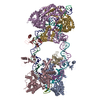
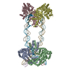
Tandem of Hermes transposase BED domain in complex with the quasi palindrome of its transposon left-end
Components
  • (Hermes transposon left-end subterminal repeats 1 and 2) x 2
  • Hermes transposase BED domain
KeywordsDNA BINDING PROTEIN/DNA / BED domain / zinc-binding domain / transposase / DNA BINDING PROTEIN-DNA complex
Function / homology
Function and homology information


nucleic acid metabolic process / protein dimerization activity / regulation of transcription by RNA polymerase II / DNA binding / identical protein binding / nucleus / metal ion binding
Similarity search - Function
Hermes trasposase, DNA-binding domain / Hermes transposase DNA-binding domain / HAT, C-terminal dimerisation domain / hAT family C-terminal dimerisation region / BED zinc finger / Zinc finger, BED-type / Zinc finger BED-type profile. / Ribonuclease H-like superfamily
Similarity search - Domain/homology
DNA / DNA (> 10) / Transposase
Similarity search - Component
Biological speciesMusca domestica (house fly)
MethodX-RAY DIFFRACTION / SYNCHROTRON / MAD / Resolution: 2.5 Å
AuthorsLannes, L. / Dyda, F.
Funding support United States, 1items
OrganizationGrant numberCountry
National Institutes of Health/National Institute of Diabetes and Digestive and Kidney Disease (NIH/NIDDK)DK036153-16 United States
CitationJournal: Nat Commun / Year: 2023
Title: Zinc-finger BED domains drive the formation of the active Hermes transpososome by asymmetric DNA binding.
Authors: Laurie Lannes / Christopher M Furman / Alison B Hickman / Fred Dyda /
Abstract: The Hermes DNA transposon is a member of the eukaryotic hAT superfamily, and its transposase forms a ring-shaped tetramer of dimers. Our investigation, combining biochemical, crystallography and cryo- ...The Hermes DNA transposon is a member of the eukaryotic hAT superfamily, and its transposase forms a ring-shaped tetramer of dimers. Our investigation, combining biochemical, crystallography and cryo-electron microscopy, and in-cell assays, shows that the full-length Hermes octamer extensively interacts with its transposon left-end through multiple BED domains of three Hermes protomers contributed by three dimers explaining the role of the unusual higher-order assembly. By contrast, the right-end is bound to no BED domains at all. Thus, this work supports a model in which Hermes multimerizes to gather enough BED domains to find its left-end among the abundant genomic DNA, facilitating the subsequent interaction with the right-end.
History
DepositionAug 30, 2022Deposition site: RCSB / Processing site: RCSB
Revision 1.0Aug 2, 2023Provider: repository / Type: Initial release
Revision 1.1May 22, 2024Group: Data collection / Category: chem_comp_atom / chem_comp_bond

-
Structure visualization

Structure viewerMolecule:
MolmilJmol/JSmol

Downloads & links

-
Assembly

Deposited unit
A: Hermes transposase BED domain
C: Hermes transposon left-end subterminal repeats 1 and 2
D: Hermes transposon left-end subterminal repeats 1 and 2
hetero molecules


Theoretical massNumber of molelcules
Total (without water)19,6284
Polymers19,5633
Non-polymers651
Water905
1
A: Hermes transposase BED domain
C: Hermes transposon left-end subterminal repeats 1 and 2
D: Hermes transposon left-end subterminal repeats 1 and 2
hetero molecules

A: Hermes transposase BED domain
C: Hermes transposon left-end subterminal repeats 1 and 2
D: Hermes transposon left-end subterminal repeats 1 and 2
hetero molecules


Theoretical massNumber of molelcules
Total (without water)39,2568
Polymers39,1256
Non-polymers1312
Water724
TypeNameSymmetry operationNumber
identity operation1_555x,y,z1
crystal symmetry operation10_665-y+1,-x+1,-z+1/61
Unit cell
Length a, b, c (Å)55.271, 55.271, 212.573
Angle α, β, γ (deg.)90.00, 90.00, 120.00
Int Tables number179
Space group name H-MP6522
Components on special symmetry positions
IDModelComponents
11C-9-

DG

-
Components

#1: Protein Hermes transposase BED domain


Mass: 9150.765 Da / Num. of mol.: 1 / Mutation: None
Source method: isolated from a genetically manipulated source
Source: (gene. exp.) Musca domestica (house fly) / Plasmid: pET15b / Details (production host): no purification tag / Production host: Escherichia coli BL21(DE3) (bacteria) / Variant (production host): Rosetta / References: UniProt: Q25442
#2: DNA chain Hermes transposon left-end subterminal repeats 1 and 2


Mass: 5201.396 Da / Num. of mol.: 1 / Mutation: None / Source method: obtained synthetically / Source: (synth.) Musca domestica (house fly)
#3: DNA chain Hermes transposon left-end subterminal repeats 1 and 2


Mass: 5210.411 Da / Num. of mol.: 1 / Source method: obtained synthetically / Source: (synth.) Musca domestica (house fly)
#4: Chemical ChemComp-ZN / ZINC ION


Mass: 65.409 Da / Num. of mol.: 1 / Source method: obtained synthetically / Formula: Zn
#5: Water ChemComp-HOH / water


Mass: 18.015 Da / Num. of mol.: 5 / Source method: isolated from a natural source / Formula: H2O
Has ligand of interestN

-
Experimental details

-
Experiment

ExperimentMethod: X-RAY DIFFRACTION / Number of used crystals: 1

-
Sample preparation

CrystalDensity Matthews: 2.46 Å3/Da / Density % sol: 50.02 %
Crystal growTemperature: 298 K / Method: vapor diffusion, hanging drop / pH: 6.5 / Details: PEG 4000, Bis-TRIS

-
Data collection

DiffractionMean temperature: 95 K / Serial crystal experiment: N
Diffraction sourceSource: SYNCHROTRON / Site: APS / Beamline: 22-ID / Wavelength: 1.28335 Å
DetectorType: DECTRIS EIGER X 16M / Detector: PIXEL / Date: Jul 31, 2020
RadiationProtocol: MAD / Monochromatic (M) / Laue (L): M / Scattering type: x-ray
Radiation wavelengthWavelength: 1.28335 Å / Relative weight: 1
ReflectionResolution: 2.5→27.64 Å / Num. obs: 7292 / % possible obs: 99.8 % / Redundancy: 19.2 % / Biso Wilson estimate: 100.25 Å2 / CC1/2: 0.999 / CC star: 1 / Rmerge(I) obs: 0.04434 / Rpim(I) all: 0.01086 / Rrim(I) all: 0.04575 / Net I/σ(I): 35.47
Reflection shellResolution: 2.5→2.6 Å / Redundancy: 19.23 % / Rmerge(I) obs: 1.224 / Mean I/σ(I) obs: 2.39 / Num. unique obs: 694 / CC1/2: 0.85 / CC star: 0.959 / Rpim(I) all: 0.2717 / Rrim(I) all: 1.224 / % possible all: 100

-
Processing

Software
NameVersionClassification
BUSTER2.10.4 (11-DEC-2020)refinement
Coot0.8.9model building
Omodel building
XDSdata reduction
XSCALEdata scaling
AutoSolphasing
RefinementMethod to determine structure: MAD / Resolution: 2.5→27.64 Å / Cor.coef. Fo:Fc: 0.94 / Cor.coef. Fo:Fc free: 0.95 / SU R Cruickshank DPI: 0.472 / Cross valid method: THROUGHOUT / σ(F): 0 / SU R Blow DPI: 0.647 / SU Rfree Blow DPI: 0.267 / SU Rfree Cruickshank DPI: 0.251
RfactorNum. reflection% reflectionSelection details
Rfree0.2382 743 10.19 %RANDOM
Rwork0.2308 ---
obs0.2316 7292 99.8 %-
Displacement parametersBiso mean: 98.38 Å2
Baniso -1Baniso -2Baniso -3
1--3.2075 Å20 Å20 Å2
2---3.2075 Å20 Å2
3---6.415 Å2
Refine analyzeLuzzati coordinate error obs: 0.35 Å
Refinement stepCycle: LAST / Resolution: 2.5→27.64 Å
ProteinNucleic acidLigandSolventTotal
Num. atoms601 691 1 5 1298
Refine LS restraints
Refine-IDTypeDev idealNumberRestraint functionWeight
X-RAY DIFFRACTIONt_bond_d0.0081425HARMONIC2
X-RAY DIFFRACTIONt_angle_deg0.942067HARMONIC2
X-RAY DIFFRACTIONt_dihedral_angle_d407SINUSOIDAL2
X-RAY DIFFRACTIONt_incorr_chiral_ct
X-RAY DIFFRACTIONt_pseud_angle
X-RAY DIFFRACTIONt_trig_c_planes
X-RAY DIFFRACTIONt_gen_planes142HARMONIC5
X-RAY DIFFRACTIONt_it1425HARMONIC5
X-RAY DIFFRACTIONt_nbd
X-RAY DIFFRACTIONt_omega_torsion3.15
X-RAY DIFFRACTIONt_other_torsion27.63
X-RAY DIFFRACTIONt_improper_torsion
X-RAY DIFFRACTIONt_chiral_improper_torsion188SEMIHARMONIC5
X-RAY DIFFRACTIONt_sum_occupancies
X-RAY DIFFRACTIONt_utility_distance4HARMONIC1
X-RAY DIFFRACTIONt_utility_angle4HARMONIC1
X-RAY DIFFRACTIONt_utility_torsion
X-RAY DIFFRACTIONt_ideal_dist_contact686SEMIHARMONIC4
LS refinement shellResolution: 2.5→2.52 Å / Total num. of bins used: 38
RfactorNum. reflection% reflection
Rfree0.3255 -7.58 %
Rwork0.3017 183 -
all0.3034 198 -
obs--100 %
Refinement TLS params.

Method: refined / Refine-ID: X-RAY DIFFRACTION

IDL112)L122)L132)L222)L232)L332)S11 (Å °)S12 (Å °)S13 (Å °)S21 (Å °)S22 (Å °)S23 (Å °)S31 (Å °)S32 (Å °)S33 (Å °)T112)T122)T132)T222)T232)T332)Origin x (Å)Origin y (Å)Origin z (Å)
16.60933.32681.39175.6090.40889.0815-0.20530.57670.7413-0.7830.0732-0.24910.31470.1640.13210.3867-0.13930.0996-0.01940.0543-0.083317.194430.17541.3939
26.02541.695-2.1899.446-2.55532.23050.4002-0.24230.1584-0.1986-0.76250.78190.0499-0.06290.36230.1693-0.36070.0212-0.02-0.0609-0.092413.160924.777218.8006
34.56312.1326-0.960311.6649-1.79167.17530.2116-0.19520.373-0.6529-0.1180.00960.0317-0.026-0.09360.0144-0.35490.0442-0.162-0.09580.11613.24524.521218.9323
Refinement TLS group
IDRefine-IDRefine TLS-IDSelection details
1X-RAY DIFFRACTION1{ A|* }
2X-RAY DIFFRACTION2{ C|* }
3X-RAY DIFFRACTION3{ D|* }

+
About Yorodumi

-
News

-
Feb 9, 2022. New format data for meta-information of EMDB entries

New format data for meta-information of EMDB entries

  • Version 3 of the EMDB header file is now the official format.
  • The previous official version 1.9 will be removed from the archive.

Related info.:EMDB header

External links:wwPDB to switch to version 3 of the EMDB data model

-
Aug 12, 2020. Covid-19 info

Covid-19 info

URL: https://pdbjlvh1.pdbj.org/emnavi/covid19.php

New page: Covid-19 featured information page in EM Navigator.

Related info.:Covid-19 info / Mar 5, 2020. Novel coronavirus structure data

+
Mar 5, 2020. Novel coronavirus structure data

Novel coronavirus structure data

Related info.:Yorodumi Speices / Aug 12, 2020. Covid-19 info

External links:COVID-19 featured content - PDBj / Molecule of the Month (242):Coronavirus Proteases

+
Jan 31, 2019. EMDB accession codes are about to change! (news from PDBe EMDB page)

EMDB accession codes are about to change! (news from PDBe EMDB page)

  • The allocation of 4 digits for EMDB accession codes will soon come to an end. Whilst these codes will remain in use, new EMDB accession codes will include an additional digit and will expand incrementally as the available range of codes is exhausted. The current 4-digit format prefixed with “EMD-” (i.e. EMD-XXXX) will advance to a 5-digit format (i.e. EMD-XXXXX), and so on. It is currently estimated that the 4-digit codes will be depleted around Spring 2019, at which point the 5-digit format will come into force.
  • The EM Navigator/Yorodumi systems omit the EMD- prefix.

Related info.:Q: What is EMD? / ID/Accession-code notation in Yorodumi/EM Navigator

External links:EMDB Accession Codes are Changing Soon! / Contact to PDBj

+
Jul 12, 2017. Major update of PDB

Major update of PDB

  • wwPDB released updated PDB data conforming to the new PDBx/mmCIF dictionary.
  • This is a major update changing the version number from 4 to 5, and with Remediation, in which all the entries are updated.
  • In this update, many items about electron microscopy experimental information are reorganized (e.g. em_software).
  • Now, EM Navigator and Yorodumi are based on the updated data.

External links:wwPDB Remediation / Enriched Model Files Conforming to OneDep Data Standards Now Available in the PDB FTP Archive

-
Yorodumi

Thousand views of thousand structures

  • Yorodumi is a browser for structure data from EMDB, PDB, SASBDB, etc.
  • This page is also the successor to EM Navigator detail page, and also detail information page/front-end page for Omokage search.
  • The word "yorodu" (or yorozu) is an old Japanese word meaning "ten thousand". "mi" (miru) is to see.

Related info.:EMDB / PDB / SASBDB / Comparison of 3 databanks / Yorodumi Search / Aug 31, 2016. New EM Navigator & Yorodumi / Yorodumi Papers / Jmol/JSmol / Function and homology information / Changes in new EM Navigator and Yorodumi

Read more