 Movie
Movie Controller

[English] 日本語
 Yorodumi
- PDB-8ea0: CryoEM structure of miniGq-coupled hM3R in complex with iperoxo (... -
+ Open data

- Basic information
| Entry | Database: PDB / ID: 8ea0 | |||||||||||||||||||||||||||||||||||||||||||||||||||||||||||||||||||||||||||||||||
|---|---|---|---|---|---|---|---|---|---|---|---|---|---|---|---|---|---|---|---|---|---|---|---|---|---|---|---|---|---|---|---|---|---|---|---|---|---|---|---|---|---|---|---|---|---|---|---|---|---|---|---|---|---|---|---|---|---|---|---|---|---|---|---|---|---|---|---|---|---|---|---|---|---|---|---|---|---|---|---|---|---|---|
| Title | CryoEM structure of miniGq-coupled hM3R in complex with iperoxo (local refinement) | |||||||||||||||||||||||||||||||||||||||||||||||||||||||||||||||||||||||||||||||||
 Components Components | Muscarinic acetylcholine receptor M3 | |||||||||||||||||||||||||||||||||||||||||||||||||||||||||||||||||||||||||||||||||
 Keywords Keywords | MEMBRANE PROTEIN / GPCR / IXO / active state / hM3R / Iperoxo | |||||||||||||||||||||||||||||||||||||||||||||||||||||||||||||||||||||||||||||||||
| Function / homology |  Function and homology information Function and homology informationsaliva secretion / phospholipase C-activating G protein-coupled acetylcholine receptor signaling pathway / Muscarinic acetylcholine receptors / Acetylcholine regulates insulin secretion / G protein-coupled acetylcholine receptor activity / phosphatidylinositol-4,5-bisphosphate phospholipase C activity / regulation of smooth muscle contraction / positive regulation of smooth muscle contraction / adenylate cyclase-inhibiting G protein-coupled acetylcholine receptor signaling pathway / acetylcholine binding ...saliva secretion / phospholipase C-activating G protein-coupled acetylcholine receptor signaling pathway / Muscarinic acetylcholine receptors / Acetylcholine regulates insulin secretion / G protein-coupled acetylcholine receptor activity / phosphatidylinositol-4,5-bisphosphate phospholipase C activity / regulation of smooth muscle contraction / positive regulation of smooth muscle contraction / adenylate cyclase-inhibiting G protein-coupled acetylcholine receptor signaling pathway / acetylcholine binding / acetylcholine receptor signaling pathway / ligand-gated ion channel signaling pathway / smooth muscle contraction / G protein-coupled receptor signaling pathway, coupled to cyclic nucleotide second messenger / basal plasma membrane / protein modification process / calcium-mediated signaling / positive regulation of insulin secretion / G protein-coupled acetylcholine receptor signaling pathway / nervous system development / signaling receptor activity / basolateral plasma membrane / G alpha (q) signalling events / chemical synaptic transmission / postsynaptic membrane / G protein-coupled receptor signaling pathway / synapse / dendrite / endoplasmic reticulum membrane / signal transduction / plasma membrane Similarity search - Function | |||||||||||||||||||||||||||||||||||||||||||||||||||||||||||||||||||||||||||||||||
| Biological species |  Homo sapiens (human) Homo sapiens (human) | |||||||||||||||||||||||||||||||||||||||||||||||||||||||||||||||||||||||||||||||||
| Method | ELECTRON MICROSCOPY / single particle reconstruction / cryo EM / Resolution: 2.56 Å | |||||||||||||||||||||||||||||||||||||||||||||||||||||||||||||||||||||||||||||||||
 Authors Authors | Zhang, S. / Fay, J.F. / Roth, B.L. | |||||||||||||||||||||||||||||||||||||||||||||||||||||||||||||||||||||||||||||||||
| Funding support |  United States, 2items United States, 2items
| |||||||||||||||||||||||||||||||||||||||||||||||||||||||||||||||||||||||||||||||||
 Citation Citation | Journal: Nature / Year: 2022 Title: Molecular basis for selective activation of DREADD-based chemogenetics. Authors: Shicheng Zhang / Ryan H Gumpper / Xi-Ping Huang / Yongfeng Liu / Brian E Krumm / Can Cao / Jonathan F Fay / Bryan L Roth / Abstract: Designer receptors exclusively activated by designer drugs (DREADDs) represent a powerful chemogenetic technology for the remote control of neuronal activity and cellular signalling. The muscarinic ...Designer receptors exclusively activated by designer drugs (DREADDs) represent a powerful chemogenetic technology for the remote control of neuronal activity and cellular signalling. The muscarinic receptor-based DREADDs are the most widely used chemogenetic tools in neuroscience research. The G-coupled DREADD (hM3Dq) is used to enhance neuronal activity, whereas the G-coupled DREADD (hM4Di) is utilized to inhibit neuronal activity. Here we report four DREADD-related cryogenic electron microscopy high-resolution structures: a hM3Dq-miniG complex and a hM4Di-miniG complex bound to deschloroclozapine; a hM3Dq-miniG complex bound to clozapine-N-oxide; and a hM3R-miniG complex bound to iperoxo. Complemented with mutagenesis, functional and computational simulation data, our structures reveal key details of the recognition of DREADD chemogenetic actuators and the molecular basis for activation. These findings should accelerate the structure-guided discovery of next-generation chemogenetic tools. | |||||||||||||||||||||||||||||||||||||||||||||||||||||||||||||||||||||||||||||||||
| History |
|
- Structure visualization
| Structure viewer | Molecule: MolmilJmol/JSmol |
|---|
- Downloads & links
-Download
| PDBx/mmCIF format | 8ea0.cif.gz | 73.9 KB | Display | PDBx/mmCIF format |
|---|---|---|---|---|
| PDB format | pdb8ea0.ent.gz | 47.3 KB | Display | PDB format |
| PDBx/mmJSON format | 8ea0.json.gz | Tree view | PDBx/mmJSON format | |
| Others |  Other downloads Other downloads |
-Validation report
| Arichive directory | https://data.pdbj.org/pub/pdb/validation_reports/ea/8ea0ftp://data.pdbj.org/pub/pdb/validation_reports/ea/8ea0 | HTTPS FTP |
|---|
-Related structure data
| Related structure data |  27970MC  8e9wC  8e9xC  8e9yC  8e9zC M: map data used to model this data C: citing same article ( |
|---|---|
| Similar structure data |


-Links
 PDBj
PDBj


- Assembly
| Deposited unit | 
|
|---|---|
| 1 |
|
-Components
| #1: Protein | Mass: 62966.617 Da / Num. of mol.: 1 Source method: isolated from a genetically manipulated source Source: (gene. exp.) Homo sapiens (human) / Gene: CHRM3 / Production host:   Spodoptera frugiperda (fall armyworm) / References: UniProt: P20309 Spodoptera frugiperda (fall armyworm) / References: UniProt: P20309 |
|---|---|
| #2: Chemical | ChemComp-IXO / 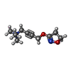  Mass: 197.254 Da / Num. of mol.: 1 / Source method: obtained synthetically / Formula: C10H17N2O2 / Feature type: SUBJECT OF INVESTIGATION Mass: 197.254 Da / Num. of mol.: 1 / Source method: obtained synthetically / Formula: C10H17N2O2 / Feature type: SUBJECT OF INVESTIGATION |
| #3: Chemical | ChemComp-Y01 /   Mass: 486.726 Da / Num. of mol.: 1 / Source method: obtained synthetically / Formula: C31H50O4 / Feature type: SUBJECT OF INVESTIGATION Mass: 486.726 Da / Num. of mol.: 1 / Source method: obtained synthetically / Formula: C31H50O4 / Feature type: SUBJECT OF INVESTIGATION |
| Has ligand of interest | Y |
| Has protein modification | Y |
-Experimental details
-Experiment
| Experiment | Method: ELECTRON MICROSCOPY |
|---|---|
| EM experiment | Aggregation state: PARTICLE / 3D reconstruction method: single particle reconstruction |
- Sample preparation
| Component | Name: hM3R / Type: COMPLEX / Entity ID: #1 / Source: RECOMBINANT |
|---|---|
| Source (natural) | Organism: Homo sapiens (human) |
| Source (recombinant) | Organism: Spodoptera frugiperda (fall armyworm) |
| Buffer solution | pH: 7.4 |
| Specimen | Embedding applied: NO / Shadowing applied: NO / Staining applied: NO / Vitrification applied: YES |
| Vitrification | Cryogen name: ETHANE-PROPANE |
- Electron microscopy imaging
| Experimental equipment |  Model: Talos Arctica / Image courtesy: FEI Company |
|---|---|
| Microscopy | Model: FEI TALOS ARCTICA |
| Electron gun | Electron source:  FIELD EMISSION GUN / Accelerating voltage: 200 kV / Illumination mode: FLOOD BEAM FIELD EMISSION GUN / Accelerating voltage: 200 kV / Illumination mode: FLOOD BEAM |
| Electron lens | Mode: BRIGHT FIELD / Nominal defocus max: 2100 nm / Nominal defocus min: 300 nm |
| Image recording | Electron dose: 59 e/Å2 / Film or detector model: GATAN K3 (6k x 4k) / Num. of real images: 2858 |
- Processing
| Software | Name: PHENIX / Version: 1.19_4092: / Classification: refinement | ||||||||||||||||||||||||
|---|---|---|---|---|---|---|---|---|---|---|---|---|---|---|---|---|---|---|---|---|---|---|---|---|---|
| EM software | Name: PHENIX / Category: model refinement | ||||||||||||||||||||||||
| CTF correction | Type: PHASE FLIPPING AND AMPLITUDE CORRECTION | ||||||||||||||||||||||||
| 3D reconstruction | Resolution: 2.56 Å / Resolution method: FSC 0.143 CUT-OFF / Num. of particles: 591814 / Symmetry type: POINT | ||||||||||||||||||||||||
| Refine LS restraints |
|
