Movie
Movie Controller


+ Open data

- Basic information
| Entry | Database: PDB / ID: 8.0E+14 | |||||||||
|---|---|---|---|---|---|---|---|---|---|---|
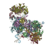
| Title | Cryo-EM structure of Rous sarcoma virus strand transfer complex | |||||||||
 Components Components |
| |||||||||
 Keywords Keywords | VIRAL PROTEIN/DNA / intasome / integrase-viral DNA complex / strand transfer complex / VIRAL PROTEIN-DNA complex | |||||||||
| Function / homology |  Function and homology information Function and homology informationHydrolases; Acting on peptide bonds (peptidases); Aspartic endopeptidases / ribonuclease H / DNA integration / viral genome integration into host DNA / establishment of integrated proviral latency / RNA-directed DNA polymerase / RNA stem-loop binding / RNA-directed DNA polymerase activity / RNA-DNA hybrid ribonuclease activity / Transferases; Transferring phosphorus-containing groups; Nucleotidyltransferases ...Hydrolases; Acting on peptide bonds (peptidases); Aspartic endopeptidases / ribonuclease H / DNA integration / viral genome integration into host DNA / establishment of integrated proviral latency / RNA-directed DNA polymerase / RNA stem-loop binding / RNA-directed DNA polymerase activity / RNA-DNA hybrid ribonuclease activity / Transferases; Transferring phosphorus-containing groups; Nucleotidyltransferases / viral nucleocapsid / DNA recombination / DNA-directed DNA polymerase / aspartic-type endopeptidase activity / Hydrolases; Acting on ester bonds / DNA-directed DNA polymerase activity / viral translational frameshifting / symbiont entry into host cell / proteolysis / DNA binding / zinc ion binding Similarity search - Function | |||||||||
| Biological species |  Rous sarcoma virus - Prague C Rous sarcoma virus - Prague C | |||||||||
| Method | ELECTRON MICROSCOPY / single particle reconstruction / cryo EM / Resolution: 3.36 Å | |||||||||
 Authors Authors | Pandey, K.K. / Bera, S. / Shi, K. / Aihara, H. / Grandgenett, D.P. | |||||||||
| Funding support |  United States, 2items United States, 2items
| |||||||||
 Citation Citation | Journal: J Biol Chem / Year: 2023 Title: Molecular determinants for Rous sarcoma virus intasome assemblies involved in retroviral integration. Authors: Sibes Bera / Ke Shi / Hideki Aihara / Duane P Grandgenett / Krishan K Pandey / Abstract: Integration of retroviral DNA into the host genome involves the formation of integrase (IN)-DNA complexes termed intasomes. Further characterization of these complexes is needed to understand their ...Integration of retroviral DNA into the host genome involves the formation of integrase (IN)-DNA complexes termed intasomes. Further characterization of these complexes is needed to understand their assembly process. Here, we report the single-particle cryo-EM structure of the Rous sarcoma virus (RSV) strand transfer complex (STC) intasome produced with IN and a preassembled viral/target DNA substrate at 3.36 Å resolution. The conserved intasome core region consisting of IN subunits contributing active sites interacting with viral/target DNA has a resolution of 3 Å. Our structure demonstrated the flexibility of the distal IN subunits relative to the IN subunits in the conserved intasome core, similar to results previously shown with the RSV octameric cleaved synaptic complex intasome produced with IN and viral DNA only. An extensive analysis of higher resolution STC structure helped in the identification of nucleoprotein interactions important for intasome assembly. Using structure-function studies, we determined the mechanisms of several IN-DNA interactions critical for assembly of both RSV intasomes. We determined the role of IN residues R244, Y246, and S124 in cleaved synaptic complex and STC intasome assemblies and their catalytic activities, demonstrating differential effects. Taken together, these studies advance our understanding of different RSV intasome structures and molecular determinants involved in their assembly. | |||||||||
| History |
|
- Structure visualization
| Structure viewer | Molecule: MolmilJmol/JSmol |
|---|
- Downloads & links
-Download
| PDBx/mmCIF format | 8e14.cif.gz | 332.3 KB | Display | PDBx/mmCIF format |
|---|---|---|---|---|
| PDB format | pdb8e14.ent.gz | 254 KB | Display | PDB format |
| PDBx/mmJSON format | 8e14.json.gz | Tree view | PDBx/mmJSON format | |
| Others |  Other downloads Other downloads |
-Validation report
| Arichive directory | https://data.pdbj.org/pub/pdb/validation_reports/e1/8e14ftp://data.pdbj.org/pub/pdb/validation_reports/e1/8e14 | HTTPS FTP |
|---|
-Related structure data
| Related structure data |  27823MC M: map data used to model this data C: citing same article ( |
|---|---|
| Similar structure data |
-Links
 PDBj
PDBj


- Assembly
| Deposited unit | 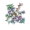
|
|---|---|
| 1 |
|
-Components
| #1: Protein | Mass: 30926.582 Da / Num. of mol.: 8 / Fragment: UNP residues 1281-1558 Source method: isolated from a genetically manipulated source Source: (gene. exp.) Rous sarcoma virus - Prague C / Strain: Prague C / Plasmid: pET11a / Production host:  References: UniProt: P03354, Hydrolases; Acting on peptide bonds (peptidases); Aspartic endopeptidases, RNA-directed DNA polymerase, DNA-directed DNA polymerase, ribonuclease H, Transferases; ...References: UniProt: P03354, Hydrolases; Acting on peptide bonds (peptidases); Aspartic endopeptidases, RNA-directed DNA polymerase, DNA-directed DNA polymerase, ribonuclease H, Transferases; Transferring phosphorus-containing groups; Nucleotidyltransferases, Hydrolases; Acting on ester bonds #2: DNA chain | Mass: 13047.456 Da / Num. of mol.: 2 / Source method: obtained synthetically / Source: (synth.) Rous sarcoma virus - Prague C#3: DNA chain | Mass: 6716.363 Da / Num. of mol.: 2 / Source method: obtained synthetically / Source: (synth.) Rous sarcoma virus - Prague C#4: DNA chain | Mass: 4821.123 Da / Num. of mol.: 2 / Source method: obtained synthetically / Source: (synth.) Rous sarcoma virus - Prague C#5: Chemical |   Mass: 65.409 Da / Num. of mol.: 2 / Source method: obtained synthetically / Formula: Zn / Feature type: SUBJECT OF INVESTIGATION Mass: 65.409 Da / Num. of mol.: 2 / Source method: obtained synthetically / Formula: Zn / Feature type: SUBJECT OF INVESTIGATIONHas ligand of interest | Y | |
|---|
-Experimental details
-Experiment
| Experiment | Method: ELECTRON MICROSCOPY |
|---|---|
| EM experiment | Aggregation state: PARTICLE / 3D reconstruction method: single particle reconstruction |
- Sample preparation
| Component | Name: RSV strand transfer complex / Type: COMPLEX / Entity ID: #1-#4 / Source: RECOMBINANT |
|---|---|
| Molecular weight | Value: 0.257 MDa / Experimental value: YES |
| Source (natural) | Organism: Rous sarcoma virus - Prague C |
| Source (recombinant) | Organism: |
| Buffer solution | pH: 7.5 |
| Specimen | Conc.: 0.5 mg/ml / Embedding applied: NO / Shadowing applied: NO / Staining applied: NO / Vitrification applied: YES |
| Specimen support | Grid material: COPPER / Grid type: Quantifoil R1.2/1.3 |
| Vitrification | Instrument: FEI VITROBOT MARK IV / Cryogen name: ETHANE / Humidity: 100 % / Chamber temperature: 277 K |
- Electron microscopy imaging
| Experimental equipment |  Model: Titan Krios / Image courtesy: FEI Company |
|---|---|
| Microscopy | Model: TFS KRIOS |
| Electron gun | Electron source:  FIELD EMISSION GUN / Accelerating voltage: 300 kV / Illumination mode: FLOOD BEAM FIELD EMISSION GUN / Accelerating voltage: 300 kV / Illumination mode: FLOOD BEAM |
| Electron lens | Mode: BRIGHT FIELD / Nominal magnification: 59000 X / Nominal defocus max: 2400 nm / Nominal defocus min: 800 nm / Cs: 0.01 mm / Alignment procedure: BASIC |
| Specimen holder | Cryogen: NITROGEN / Specimen holder model: FEI TITAN KRIOS AUTOGRID HOLDER |
| Image recording | Electron dose: 50 e/Å2 / Detector mode: SUPER-RESOLUTION / Film or detector model: FEI FALCON IV (4k x 4k) / Num. of real images: 3000 |
| EM imaging optics | Energyfilter name: GIF Bioquantum / Energyfilter slit width: 20 eV |
| Image scans | Movie frames/image: 40 |
- Processing
| Software | Name: PHENIX / Version: 1.20.1_4487: / Classification: refinement | ||||||||||||||||||||||||
|---|---|---|---|---|---|---|---|---|---|---|---|---|---|---|---|---|---|---|---|---|---|---|---|---|---|
| EM software |
| ||||||||||||||||||||||||
| CTF correction | Type: NONE | ||||||||||||||||||||||||
| Particle selection | Num. of particles selected: 1811357 | ||||||||||||||||||||||||
| 3D reconstruction | Resolution: 3.36 Å / Resolution method: FSC 0.143 CUT-OFF / Num. of particles: 141000 / Symmetry type: POINT | ||||||||||||||||||||||||
| Atomic model building | B value: 30 / Protocol: RIGID BODY FIT / Space: REAL / Target criteria: correlation coefficient | ||||||||||||||||||||||||
| Atomic model building | PDB-ID: 5EJK Accession code: 5EJK / Source name: PDB / Type: experimental model | ||||||||||||||||||||||||
| Refine LS restraints |
|