National Institutes of Health/National Institute of General Medical Sciences (NIH/NIGMS)
GM141553
United States
National Institutes of Health/National Cancer Institute (NIH/NCI)
CA008748
United States
Citation
Journal: Proc Natl Acad Sci U S A / Year: 2022 Title: Mechanism of 4-aminopyridine inhibition of the lysosomal channel TMEM175. Authors: SeCheol Oh / Robyn Stix / Wenchang Zhou / José D Faraldo-Gómez / Richard K Hite / Abstract: Transmembrane protein 175 (TMEM175) is an evolutionarily distinct lysosomal cation channel whose mutation is associated with the development of Parkinson's disease. Here, we present a cryoelectron ...Transmembrane protein 175 (TMEM175) is an evolutionarily distinct lysosomal cation channel whose mutation is associated with the development of Parkinson's disease. Here, we present a cryoelectron microscopy structure and molecular simulations of TMEM175 bound to 4-aminopyridine (4-AP), the only known small-molecule inhibitor of TMEM175 and a broad K channel inhibitor, as well as a drug approved by the Food and Drug Administration against multiple sclerosis. The structure shows that 4-AP, whose mode of action had not been previously visualized, binds near the center of the ion conduction pathway, in the open state of the channel. Molecular dynamics simulations reveal that this binding site is near the middle of the transmembrane potential gradient, providing a rationale for the voltage-dependent dissociation of 4-AP from TMEM175. Interestingly, bound 4-AP rapidly switches between three predominant binding poses, stabilized by alternate interaction patterns dictated by the twofold symmetry of the channel. Despite this highly dynamic binding mode, bound 4-AP prevents not only ion permeation but also water flow. Together, these studies provide a framework for the rational design of novel small-molecule inhibitors of TMEM175 that might reveal the role of this channel in human lysosomal physiology both in health and disease.
Mass: 55667.219 Da / Num. of mol.: 2 Source method: isolated from a genetically manipulated source Source: (gene. exp.) Homo sapiens (human) / Gene: TMEM175 / Production host: Homo sapiens (human) / References: UniProt: Q9BSA9
In the structure databanks used in Yorodumi, some data are registered as the other names, "COVID-19 virus" and "2019-nCoV". Here are the details of the virus and the list of structure data.
Jan 31, 2019. EMDB accession codes are about to change! (news from PDBe EMDB page)
EMDB accession codes are about to change! (news from PDBe EMDB page)
The allocation of 4 digits for EMDB accession codes will soon come to an end. Whilst these codes will remain in use, new EMDB accession codes will include an additional digit and will expand incrementally as the available range of codes is exhausted. The current 4-digit format prefixed with “EMD-” (i.e. EMD-XXXX) will advance to a 5-digit format (i.e. EMD-XXXXX), and so on. It is currently estimated that the 4-digit codes will be depleted around Spring 2019, at which point the 5-digit format will come into force.
The EM Navigator/Yorodumi systems omit the EMD- prefix.
Related info.:Q: What is EMD? / ID/Accession-code notation in Yorodumi/EM Navigator
Yorodumi is a browser for structure data from EMDB, PDB, SASBDB, etc.
This page is also the successor to EM Navigator detail page, and also detail information page/front-end page for Omokage search.
The word "yorodu" (or yorozu) is an old Japanese word meaning "ten thousand". "mi" (miru) is to see.
Related info.:EMDB / PDB / SASBDB / Comparison of 3 databanks / Yorodumi Search / Aug 31, 2016. New EM Navigator & Yorodumi / Yorodumi Papers / Jmol/JSmol / Function and homology information / Changes in new EM Navigator and Yorodumi
 Movie
Movie Controller


 Open data
 Basic information
 Components
 Keywords
 Function and homology information
 Homo sapiens (human)
 Authors
 United States, 2items
  Citation
 Structure visualization
 Downloads & links
 Other downloads
 PDBj
PDBj
 Assembly
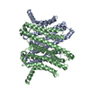

 Mass: 39.098 Da / Num. of mol.: 4 / Source method: obtained synthetically / Formula: K
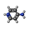
 Mass: 95.122 Da / Num. of mol.: 1 / Source method: obtained synthetically / Formula: C5H7N2 / Feature type: SUBJECT OF INVESTIGATION
 Mass: 18.015 Da / Num. of mol.: 91 / Source method: isolated from a natural source / Formula: H2O
 Sample preparation
 Electron microscopy imaging
 FIELD EMISSION GUN / Accelerating voltage: 300 kV / Illumination mode: FLOOD BEAM
 Processing
