 Movie
Movie Controller

[English] 日本語
 Yorodumi
- PDB-8cr2: Homo sapiens Get1/Get2 heterotetramer (a3' deletion variant) in c... -
+ Open data

- Basic information
| Entry | Database: PDB / ID: 8cr2 | |||||||||
|---|---|---|---|---|---|---|---|---|---|---|
| Title | Homo sapiens Get1/Get2 heterotetramer (a3' deletion variant) in complex with a Get3 dimer | |||||||||
 Components Components |
| |||||||||
 Keywords Keywords | MEMBRANE PROTEIN / membrane protein insertion / GET pathway / tail anchored membrane protein | |||||||||
| Function / homology |  Function and homology information Function and homology informationotic vesicle development / GET complex / protein carrier activity / tail-anchored membrane protein insertion into ER membrane / receptor recycling / protein insertion into ER membrane / post-translational protein targeting to endoplasmic reticulum membrane / Insertion of tail-anchored proteins into the endoplasmic reticulum membrane / B cell homeostasis / vesicle-mediated transport ...otic vesicle development / GET complex / protein carrier activity / tail-anchored membrane protein insertion into ER membrane / receptor recycling / protein insertion into ER membrane / post-translational protein targeting to endoplasmic reticulum membrane / Insertion of tail-anchored proteins into the endoplasmic reticulum membrane / B cell homeostasis / vesicle-mediated transport / protein-membrane adaptor activity / negative regulation of protein ubiquitination / negative regulation of proteasomal ubiquitin-dependent protein catabolic process / establishment of localization in cell / defense response / sensory perception of sound / synapse organization / Hydrolases; Acting on acid anhydrides; Acting on acid anhydrides to facilitate cellular and subcellular movement / epidermal growth factor receptor signaling pathway / protein stabilization / ubiquitin protein ligase binding / endoplasmic reticulum membrane / nucleolus / endoplasmic reticulum / signal transduction / ATP hydrolysis activity / extracellular exosome / nucleoplasm / ATP binding / membrane / metal ion binding / nucleus / cytoplasm Similarity search - Function | |||||||||
| Biological species |  Homo sapiens (human) Homo sapiens (human) | |||||||||
| Method | ELECTRON MICROSCOPY / single particle reconstruction / cryo EM / Resolution: 4.2 Å | |||||||||
 Authors Authors | McDowell, M.A. / Heimes, M. / Wild, K. / Sinning, I. | |||||||||
| Funding support |  Germany, 2items Germany, 2items
| |||||||||
 Citation Citation | Journal: Nat Commun / Year: 2023 Title: The GET insertase exhibits conformational plasticity and induces membrane thinning. Authors: Melanie A McDowell / Michael Heimes / Giray Enkavi / Ákos Farkas / Daniel Saar / Klemens Wild / Blanche Schwappach / Ilpo Vattulainen / Irmgard Sinning /  Abstract: The eukaryotic guided entry of tail-anchored proteins (GET) pathway mediates the biogenesis of tail-anchored (TA) membrane proteins at the endoplasmic reticulum. In the cytosol, the Get3 chaperone ...The eukaryotic guided entry of tail-anchored proteins (GET) pathway mediates the biogenesis of tail-anchored (TA) membrane proteins at the endoplasmic reticulum. In the cytosol, the Get3 chaperone captures the TA protein substrate and delivers it to the Get1/Get2 membrane protein complex (GET insertase), which then inserts the substrate via a membrane-embedded hydrophilic groove. Here, we present structures, atomistic simulations and functional data of human and Chaetomium thermophilum Get1/Get2/Get3. The core fold of the GET insertase is conserved throughout eukaryotes, whilst thinning of the lipid bilayer occurs in the vicinity of the hydrophilic groove to presumably lower the energetic barrier of membrane insertion. We show that the gating interaction between Get2 helix α3' and Get3 drives conformational changes in both Get3 and the Get1/Get2 membrane heterotetramer. Thus, we provide a framework to understand the conformational plasticity of the GET insertase and how it remodels its membrane environment to promote substrate insertion. | |||||||||
| History |
|
- Structure visualization
| Structure viewer | Molecule: MolmilJmol/JSmol |
|---|
- Downloads & links
-Download
| PDBx/mmCIF format | 8cr2.cif.gz | 173.6 KB | Display | PDBx/mmCIF format |
|---|---|---|---|---|
| PDB format | pdb8cr2.ent.gz | 130.9 KB | Display | PDB format |
| PDBx/mmJSON format | 8cr2.json.gz | Tree view | PDBx/mmJSON format | |
| Others |  Other downloads Other downloads |
-Validation report
| Arichive directory | https://data.pdbj.org/pub/pdb/validation_reports/cr/8cr2ftp://data.pdbj.org/pub/pdb/validation_reports/cr/8cr2 | HTTPS FTP |
|---|
-Related structure data
| Related structure data |  16802MC  8cqzC  8cr1C  8oduC  8odvC M: map data used to model this data C: citing same article ( |
|---|---|
| Similar structure data |


-Links
 PDBj
PDBj


- Assembly
| Deposited unit | 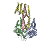
|
|---|---|
| 1 |
|
-Components
| #1: Protein | Mass: 40146.070 Da / Num. of mol.: 2 Source method: isolated from a genetically manipulated source Details: Get3 was expressed in E. coli / Source: (gene. exp.) Homo sapiens (human) / Gene: ASNA1, ARSA, TRC40 / Production host:  References: UniProt: O43681, Hydrolases; Acting on acid anhydrides #2: Protein | Mass: 34429.418 Da / Num. of mol.: 2 Mutation: Truncation of 185 N-terminal residues. Residues 242-250 replaced with a GGGG linker Source method: isolated from a genetically manipulated source Details: Get2-Get1 was expressed as a fusion protein in S. frugiperda Source: (gene. exp.) Homo sapiens (human) / Gene: CAMLG, CAML, GET2, GET1, CHD5, WRB / Production host:   Spodoptera frugiperda (fall armyworm) / References: UniProt: P49069, UniProt: O00258 Spodoptera frugiperda (fall armyworm) / References: UniProt: P49069, UniProt: O00258#3: Chemical | ChemComp-ZN / |   Mass: 65.409 Da / Num. of mol.: 1 / Source method: obtained synthetically / Formula: Zn Mass: 65.409 Da / Num. of mol.: 1 / Source method: obtained synthetically / Formula: ZnHas ligand of interest | N | Has protein modification | Y | |
|---|
-Experimental details
-Experiment
| Experiment | Method: ELECTRON MICROSCOPY |
|---|---|
| EM experiment | Aggregation state: PARTICLE / 3D reconstruction method: single particle reconstruction |
- Sample preparation
| Component |
| ||||||||||||||||||||||||||||
|---|---|---|---|---|---|---|---|---|---|---|---|---|---|---|---|---|---|---|---|---|---|---|---|---|---|---|---|---|---|
| Molecular weight |
| ||||||||||||||||||||||||||||
| Source (natural) |
| ||||||||||||||||||||||||||||
| Source (recombinant) |
| ||||||||||||||||||||||||||||
| Buffer solution | pH: 7.5 | ||||||||||||||||||||||||||||
| Buffer component |
| ||||||||||||||||||||||||||||
| Specimen | Conc.: 1.2 mg/ml / Embedding applied: NO / Shadowing applied: NO / Staining applied: NO / Vitrification applied: YES / Details: Complex stabilised in PMAL-C8 amphipol | ||||||||||||||||||||||||||||
| Specimen support | Grid material: COPPER / Grid mesh size: 300 divisions/in. / Grid type: Quantifoil R2/1 | ||||||||||||||||||||||||||||
| Vitrification | Instrument: FEI VITROBOT MARK IV / Cryogen name: ETHANE / Humidity: 95 % / Chamber temperature: 279 K |
- Electron microscopy imaging
| Experimental equipment |  Model: Titan Krios / Image courtesy: FEI Company |
|---|---|
| Microscopy | Model: FEI TITAN KRIOS |
| Electron gun | Electron source:  FIELD EMISSION GUN / Accelerating voltage: 300 kV / Illumination mode: FLOOD BEAM FIELD EMISSION GUN / Accelerating voltage: 300 kV / Illumination mode: FLOOD BEAM |
| Electron lens | Mode: BRIGHT FIELD / Nominal defocus max: 2400 nm / Nominal defocus min: 1200 nm / Cs: 2.7 mm |
| Specimen holder | Cryogen: NITROGEN |
| Image recording | Average exposure time: 2.58 sec. / Electron dose: 53.2 e/Å2 / Detector mode: COUNTING / Film or detector model: GATAN K3 (6k x 4k) / Num. of grids imaged: 1 / Num. of real images: 12311 |
- Processing
| Software | Name: PHENIX / Version: 1.19_4092: / Classification: refinement | ||||||||||||||||||||||||||||||||||||
|---|---|---|---|---|---|---|---|---|---|---|---|---|---|---|---|---|---|---|---|---|---|---|---|---|---|---|---|---|---|---|---|---|---|---|---|---|---|
| EM software |
| ||||||||||||||||||||||||||||||||||||
| CTF correction | Type: PHASE FLIPPING AND AMPLITUDE CORRECTION | ||||||||||||||||||||||||||||||||||||
| Particle selection | Num. of particles selected: 2044854 | ||||||||||||||||||||||||||||||||||||
| Symmetry | Point symmetry: C1 (asymmetric) | ||||||||||||||||||||||||||||||||||||
| 3D reconstruction | Resolution: 4.2 Å / Resolution method: FSC 0.143 CUT-OFF / Num. of particles: 224354 / Symmetry type: POINT | ||||||||||||||||||||||||||||||||||||
| Atomic model building | Protocol: RIGID BODY FIT / Space: REAL | ||||||||||||||||||||||||||||||||||||
| Atomic model building | PDB-ID: 6SO5 Accession code: 6SO5 / Source name: PDB / Type: experimental model | ||||||||||||||||||||||||||||||||||||
| Refine LS restraints |
|

