 Movie
Movie Controller

[English] 日本語
 Yorodumi
- PDB-8bcp: Cryo-EM structure of the proximal end of bacteriophage T5 tail : ... -
+ Open data

- Basic information
| Entry | Database: PDB / ID: 8bcp | |||||||||
|---|---|---|---|---|---|---|---|---|---|---|
| Title | Cryo-EM structure of the proximal end of bacteriophage T5 tail : p142 tail terminator protein hexamer and pb6 tail tube protein trimer | |||||||||
 Components Components |
| |||||||||
 Keywords Keywords | VIRAL PROTEIN / Bacteriophage / Siphophage / T5 / tail terminator / Trp | |||||||||
| Function / homology |  Function and homology information Function and homology informationvirus tail, tube / symbiont genome ejection through host cell envelope, long flexible tail mechanism / viral tail assembly / virus tail / viral release from host cell by cytolysis Similarity search - Function | |||||||||
| Biological species |  Escherichia phage T5 (virus) Escherichia phage T5 (virus) | |||||||||
| Method | ELECTRON MICROSCOPY / single particle reconstruction / cryo EM / Resolution: 3.88 Å | |||||||||
 Authors Authors | Linares, R. / Effantin, G. / Breyton, C. | |||||||||
| Funding support |  France, 2items France, 2items
| |||||||||
 Citation Citation | Journal: J Virol / Year: 2025 Title: About bacteriophage tail terminator and tail completion proteins: structure of the proximal extremity of siphophage T5 tail. Authors: Romain Linares / Cécile Breyton / Abstract: Bacteriophages are viruses infecting bacteria. The vast majority of them bear a tail, allowing host recognition, cell wall perforation, and DNA injection into the host cytoplasm. Using electron cryo- ...Bacteriophages are viruses infecting bacteria. The vast majority of them bear a tail, allowing host recognition, cell wall perforation, and DNA injection into the host cytoplasm. Using electron cryo-microscopy (cryo-EM) and single particle analysis, we determined the organization of the tail proximal extremity of siphophage T5 that possesses a long flexible tail and solved the structure of its tail terminator protein p142 (TrP). It allowed us to confirm the common evolutionary origin between T5 TrP and other known or putative TrPs from siphophages, myophages, and bacterial tail-like machines, despite very poor sequence conservation. By also determining the structure of the T5 tail proximal extremity after interaction with T5 bacterial receptor FhuA, we showed that no conformational changes occur in TrP and confirmed that the infection signal transduction is not carried by the tube itself. We also investigated the location of T5 Neck1 or tail completion protein p143 (TCP) and showed, thanks to a combination of cryo-EM and structure prediction using Alphafold2, that it is not located at the capsid-to-tail interface as suggested by its position in the genome, but instead, very unexpectedly, on the side of T5 tail tip, and that it appears to be monomeric. Based on structure comparison with other putative TCPs predicted structures, this feature could not be shared by other TCPs and questions the affiliation of p143 to this family of protein.IMPORTANCEBacteriophages, viruses infecting bacteria, are the most abundant living entities on Earth. They are present in all ecosystems where bacteria develop and are instrumental in the regulation, diversity, evolution, and pathogeny of microbial populations. Moreover, with the increasing number of pathogenic strains resistant to antibiotics, virulent phages are considered a serious alternative or complement to classical treatments. 96% of all phages present a tail that allows host recognition and safe channeling of the DNA to the host cytoplasm. We present the atomic model of the proximal extremity of the siphophage T5 tail, confirming structural similarities with other phages. This structure, combined with results previously published and further explored, also allowed a review and a discussion on the role and localization of a mysterious tail protein, the tail completion protein, which is known to be present in the phage tails, but that was never identified in a phage structure. | |||||||||
| History |
|
- Structure visualization
| Structure viewer | Molecule: MolmilJmol/JSmol |
|---|
- Downloads & links
-Download
| PDBx/mmCIF format | 8bcp.cif.gz | 395.9 KB | Display | PDBx/mmCIF format |
|---|---|---|---|---|
| PDB format | pdb8bcp.ent.gz | Display | PDB format | |
| PDBx/mmJSON format | 8bcp.json.gz | Tree view | PDBx/mmJSON format | |
| Others |  Other downloads Other downloads |
-Validation report
| Arichive directory | https://data.pdbj.org/pub/pdb/validation_reports/bc/8bcpftp://data.pdbj.org/pub/pdb/validation_reports/bc/8bcp | HTTPS FTP |
|---|
-Related structure data
| Related structure data |  15967MC 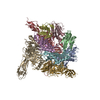 8bcuC M: map data used to model this data C: citing same article ( |
|---|---|
| Similar structure data |
-Links
 PDBj
PDBj- Assembly
| Deposited unit | 
|
|---|---|
| 1 |
|
-Components
| #1: Protein | Mass: 18378.643 Da / Num. of mol.: 6 / Source method: isolated from a natural source / Source: (natural) Escherichia phage T5 (virus) / References: UniProt: Q6QGE1#2: Protein | Mass: 50459.215 Da / Num. of mol.: 3 / Source method: isolated from a natural source / Source: (natural) Escherichia phage T5 (virus) / References: UniProt: Q6QGE2Has protein modification | N | |
|---|
-Experimental details
-Experiment
| Experiment | Method: ELECTRON MICROSCOPY |
|---|---|
| EM experiment | Aggregation state: PARTICLE / 3D reconstruction method: single particle reconstruction |
- Sample preparation
| Component | Name: Escherichia virus T5 / Type: VIRUS Details: Pure T5 tails obtained by infecting E. coli F strain with the amber mutant phage T5D20am30d Entity ID: all / Source: NATURAL | |||||||||||||||||||||||||
|---|---|---|---|---|---|---|---|---|---|---|---|---|---|---|---|---|---|---|---|---|---|---|---|---|---|---|
| Molecular weight | Experimental value: NO | |||||||||||||||||||||||||
| Source (natural) | Organism: Escherichia virus T5 / Strain: T5D20am30d | |||||||||||||||||||||||||
| Details of virus | Empty: NO / Enveloped: NO / Isolate: STRAIN / Type: VIRUS-LIKE PARTICLE | |||||||||||||||||||||||||
| Natural host | Organism: Escherichia coli / Strain: F | |||||||||||||||||||||||||
| Buffer solution | pH: 8 | |||||||||||||||||||||||||
| Buffer component |
| |||||||||||||||||||||||||
| Specimen | Embedding applied: NO / Shadowing applied: NO / Staining applied: NO / Vitrification applied: YES Details: Pure T5 tails obtained by infecting E. coli F strain with the amber mutant phage T5D20am30d | |||||||||||||||||||||||||
| Specimen support | Details: 25 mA / Grid material: COPPER/RHODIUM / Grid mesh size: 300 divisions/in. / Grid type: Quantifoil R2/1 | |||||||||||||||||||||||||
| Vitrification | Instrument: FEI VITROBOT MARK IV / Cryogen name: ETHANE / Humidity: 100 % / Chamber temperature: 293.15 K Details: 3 uL of T5 tails sample were deposited on a freshly glow discharged EM grid and plunge-frozen in nitrogen-cooled liquid ethane using a ThermoFisher Mark IV Vitrobot device (100 percent ...Details: 3 uL of T5 tails sample were deposited on a freshly glow discharged EM grid and plunge-frozen in nitrogen-cooled liquid ethane using a ThermoFisher Mark IV Vitrobot device (100 percent humidity, 20 Celsius degrees, 5 s blotting time, blot force 0) |
- Electron microscopy imaging
| Experimental equipment |  Model: Titan Krios / Image courtesy: FEI Company |
|---|---|
| Microscopy | Model: FEI TITAN KRIOS |
| Electron gun | Electron source:  FIELD EMISSION GUN / Accelerating voltage: 300 kV / Illumination mode: FLOOD BEAM FIELD EMISSION GUN / Accelerating voltage: 300 kV / Illumination mode: FLOOD BEAM |
| Electron lens | Mode: BRIGHT FIELD / Nominal magnification: 105000 X / Nominal defocus max: 3000 nm / Nominal defocus min: 1000 nm / Cs: 2.7 mm |
| Specimen holder | Cryogen: NITROGEN / Specimen holder model: FEI TITAN KRIOS AUTOGRID HOLDER |
| Image recording | Electron dose: 40 e/Å2 / Detector mode: COUNTING / Film or detector model: GATAN K2 SUMMIT (4k x 4k) |
| Image scans | Movie frames/image: 40 |
- Processing
| Software | Name: PHENIX / Version: 1.20.1_4487: / Classification: refinement | ||||||||||||||||||||||||||||||||
|---|---|---|---|---|---|---|---|---|---|---|---|---|---|---|---|---|---|---|---|---|---|---|---|---|---|---|---|---|---|---|---|---|---|
| EM software |
| ||||||||||||||||||||||||||||||||
| CTF correction | Type: PHASE FLIPPING AND AMPLITUDE CORRECTION | ||||||||||||||||||||||||||||||||
| Symmetry | Point symmetry: C3 (3 fold cyclic) | ||||||||||||||||||||||||||||||||
| 3D reconstruction | Resolution: 3.88 Å / Resolution method: FSC 0.143 CUT-OFF / Num. of particles: 9952 / Symmetry type: POINT | ||||||||||||||||||||||||||||||||
| Atomic model building | B value: 146 / Protocol: BACKBONE TRACE / Space: REAL | ||||||||||||||||||||||||||||||||
| Refine LS restraints |
|
