Defective VWF binding to collagen type I / Enhanced cleavage of VWF variant by ADAMTS13 / Defective VWF cleavage by ADAMTS13 variant / Weibel-Palade body / Defective F8 binding to von Willebrand factor / Enhanced binding of GP1BA variant to VWF multimer:collagen / Defective binding of VWF variant to GPIb:IX:V / hemostasis / platelet alpha granule / Platelet Adhesion to exposed collagen ...Defective VWF binding to collagen type I / Enhanced cleavage of VWF variant by ADAMTS13 / Defective VWF cleavage by ADAMTS13 variant / Weibel-Palade body / Defective F8 binding to von Willebrand factor / Enhanced binding of GP1BA variant to VWF multimer:collagen / Defective binding of VWF variant to GPIb:IX:V / hemostasis / platelet alpha granule / Platelet Adhesion to exposed collagen / positive regulation of intracellular signal transduction / GP1b-IX-V activation signalling / p130Cas linkage to MAPK signaling for integrins / cell-substrate adhesion / Defective F8 cleavage by thrombin / Platelet Aggregation (Plug Formation) / immunoglobulin binding / GRB2:SOS provides linkage to MAPK signaling for Integrins / Integrin cell surface interactions / collagen binding / Intrinsic Pathway of Fibrin Clot Formation / Integrin signaling / extracellular matrix / platelet alpha granule lumen / Signaling by high-kinase activity BRAF mutants / MAP2K and MAPK activation / platelet activation / response to wounding / Signaling by RAF1 mutants / Signaling by moderate kinase activity BRAF mutants / Paradoxical activation of RAF signaling by kinase inactive BRAF / Signaling downstream of RAS mutants / Signaling by BRAF and RAF1 fusions / blood coagulation / integrin binding / Platelet degranulation / protein-folding chaperone binding / protease binding / collagen-containing extracellular matrix / cell adhesion / endoplasmic reticulum / extracellular space / extracellular exosome / extracellular region / identical protein binding 類似検索 - 分子機能
von Willebrand factor, VWA N-terminal domain / Von Willebrand factor / VWA N-terminal / C8 domain / Uncharacterised domain, cysteine-rich / C8 / von Willebrand factor, type D domain / von Willebrand factor type D domain / VWFD domain profile. / von Willebrand factor (vWF) type D domain ...von Willebrand factor, VWA N-terminal domain / Von Willebrand factor / VWA N-terminal / C8 domain / Uncharacterised domain, cysteine-rich / C8 / von Willebrand factor, type D domain / von Willebrand factor type D domain / VWFD domain profile. / von Willebrand factor (vWF) type D domain / C-terminal cystine knot signature. / von Willebrand factor (vWF) type C domain / Trypsin Inhibitor-like, cysteine rich domain / Serine protease inhibitor-like superfamily / Trypsin Inhibitor like cysteine rich domain / C-terminal cystine knot domain profile. / Cystine knot, C-terminal / C-terminal cystine knot-like domain (CTCK) / von Willebrand factor type C domain / VWFC domain signature. / VWFC domain profile. / von Willebrand factor (vWF) type C domain / VWFC domain / von Willebrand factor type A domain / von Willebrand factor (vWF) type A domain / VWFA domain profile. / von Willebrand factor, type A / von Willebrand factor A-like domain superfamily 類似検索 - ドメイン・相同性
ジャーナル: Blood / 年: 2022 タイトル: Assembly of von Willebrand factor tubules with in vivo helical parameters requires A1 domain insertion. 著者: Gabriel Javitt / Noa Yeshaya / Lev Khmelnitsky / Deborah Fass / 要旨: The von Willebrand factor (VWF) glycoprotein is stored in tubular form in Weibel-Palade bodies (WPBs) before secretion from endothelial cells into the bloodstream. The organization of VWF in the ...The von Willebrand factor (VWF) glycoprotein is stored in tubular form in Weibel-Palade bodies (WPBs) before secretion from endothelial cells into the bloodstream. The organization of VWF in the tubules promotes formation of covalently linked VWF polymers and enables orderly secretion without polymer tangling. Recent studies have described the high-resolution structure of helical tubular cores formed in vitro by the D1D2 and D'D3 amino-terminal protein segments of VWF. Here we show that formation of tubules with the helical geometry observed for VWF in intracellular WPBs requires also the VWA1 (A1) domain. We reconstituted VWF tubules from segments containing the A1 domain and discovered it to be inserted between helical turns of the tubule, altering helical parameters and explaining the increased robustness of tubule formation when A1 is present. The conclusion from this observation is that the A1 domain has a direct role in VWF assembly, along with its known activity in hemostasis after secretion.
 ムービー
ムービー コントローラー


 データを開く
 基本情報
基本情報 要素
要素 キーワード
キーワード 機能・相同性情報
 Homo sapiens (ヒト)
 データ登録者
 イスラエル, 1件
  引用
引用 構造の表示
構造の表示 ダウンロードとリンク
 その他のダウンロード


 PDBj
PDBj


 集合体
集合体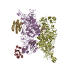

 タイプ: D-saccharide, beta linking / 分子量: 221.208 Da / 分子数: 8 / 由来タイプ: 合成 / 式: C8H15NO6
 分子量: 40.078 Da / 分子数: 6 / 由来タイプ: 合成 / 式: Ca
 試料調製
試料調製 電子顕微鏡撮影
 FIELD EMISSION GUN / 加速電圧: 300 kV / 照射モード: FLOOD BEAM
 解析
解析