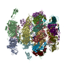
 ムービー
ムービー コントローラー


+ データを開く

- 基本情報
基本情報
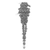
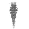
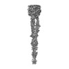
| 登録情報 | データベース: PDB / ID: 7zqb | |||||||||
|---|---|---|---|---|---|---|---|---|---|---|
| タイトル | Tail tip of siphophage T5 : full structure | |||||||||
 要素 要素 |
| |||||||||
 キーワード キーワード | VIRAL PROTEIN / Bacteriophage / Siphophage / T5 / baseplate | |||||||||
| 機能・相同性 |  機能・相同性情報 機能・相同性情報peptidoglycan muralytic activity / : / symbiont entry into host cell via disruption of host cell wall peptidoglycan / virus tail, tube / symbiont genome ejection through host cell envelope, long flexible tail mechanism / virus tail, baseplate / viral tail assembly / virus tail, fiber / symbiont entry into host cell via disruption of host cell envelope / virus tail ...peptidoglycan muralytic activity / : / symbiont entry into host cell via disruption of host cell wall peptidoglycan / virus tail, tube / symbiont genome ejection through host cell envelope, long flexible tail mechanism / virus tail, baseplate / viral tail assembly / virus tail, fiber / symbiont entry into host cell via disruption of host cell envelope / virus tail / symbiont entry into host / metabolic process / viral release from host cell by cytolysis / symbiont genome entry into host cell via pore formation in plasma membrane / killing of cells of another organism / lyase activity / defense response to bacterium 類似検索 - 分子機能 | |||||||||
| 生物種 |  Escherichia phage T5 (ファージ) Escherichia phage T5 (ファージ) | |||||||||
| 手法 | 電子顕微鏡法 / 単粒子再構成法 / クライオ電子顕微鏡法 / 解像度: 3.88 Å | |||||||||
 データ登録者 データ登録者 | Linares, R. / Arnaud, C.A. / Effantin, G. / Darnault, C. / Epalle, N. / Boeri Erba, E. / Schoehn, G. / Breyton, C. | |||||||||
| 資金援助 |  フランス, 2件 フランス, 2件
| |||||||||
 引用 引用 | ジャーナル: Sci Adv / 年: 2023 タイトル: Structural basis of bacteriophage T5 infection trigger and cell wall perforation. 著者: Romain Linares / Charles-Adrien Arnaud / Grégory Effantin / Claudine Darnault / Nathan Hugo Epalle / Elisabetta Boeri Erba / Guy Schoehn / Cécile Breyton / 要旨: Most bacteriophages present a tail allowing host recognition, cell wall perforation, and viral DNA channeling from the capsid to the infected bacterium cytoplasm. The majority of tailed phages bear a ...Most bacteriophages present a tail allowing host recognition, cell wall perforation, and viral DNA channeling from the capsid to the infected bacterium cytoplasm. The majority of tailed phages bear a long flexible tail () at the tip of which receptor binding proteins (RBPs) specifically interact with their host, triggering infection. In siphophage T5, the unique RBP is located at the extremity of a central fiber. We present the structures of T5 tail tip, determined by cryo-electron microscopy before and after interaction with its receptor, FhuA, reconstituted into nanodisc. These structures bring out the important conformational changes undergone by T5 tail tip upon infection, which include bending of T5 central fiber on the side of the tail tip, tail anchoring to the membrane, tail tube opening, and formation of a transmembrane channel. The data allow to detail the first steps of an otherwise undescribed infection mechanism. | |||||||||
| 履歴 |
|
- 構造の表示
構造の表示
| 構造ビューア | 分子: MolmilJmol/JSmol |
|---|
- ダウンロードとリンク
-ダウンロード
| PDBx/mmCIF形式 | 7zqb.cif.gz | 1.9 MB | 表示 | PDBx/mmCIF形式 |
|---|---|---|---|---|
| PDB形式 | pdb7zqb.ent.gz | 表示 | PDB形式 | |
| PDBx/mmJSON形式 | 7zqb.json.gz | ツリー表示 | PDBx/mmJSON形式 | |
| その他 |  その他のダウンロード その他のダウンロード |
-検証レポート
| 文書・要旨 | 7zqb_validation.pdf.gz | 1.2 MB | 表示 | wwPDB検証レポート |
|---|---|---|---|---|
| 文書・詳細版 | 7zqb_full_validation.pdf.gz | 1.2 MB | 表示 | |
| XML形式データ | 7zqb_validation.xml.gz | 252.7 KB | 表示 | |
| CIF形式データ | 7zqb_validation.cif.gz | 400.7 KB | 表示 | |
| アーカイブディレクトリ | https://data.pdbj.org/pub/pdb/validation_reports/zq/7zqbftp://data.pdbj.org/pub/pdb/validation_reports/zq/7zqb | HTTPS FTP |
-関連構造データ


-リンク
 PDBj
PDBj- 集合体
集合体
| 登録構造単位 | 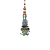
|
|---|---|
| 1 |
|
-要素
-タンパク質 , 7種, 36分子 ihjRQSNMOJLUKPTIHGaXYWVZFECDAB...
| #1: タンパク質 | 分子量: 74851.703 Da / 分子数: 3 / 由来タイプ: 天然 / 由来: (天然) Escherichia phage T5 (ファージ) / 参照: UniProt: Q6QGF0#2: タンパク質 | 分子量: 15079.864 Da / 分子数: 12 / 由来タイプ: 天然 / 由来: (天然) Escherichia phage T5 (ファージ) / 参照: UniProt: Q7Y5D9#3: タンパク質 | 分子量: 34367.605 Da / 分子数: 3 / 由来タイプ: 天然 / 由来: (天然) Escherichia phage T5 (ファージ) / 参照: UniProt: Q6QGE3#4: タンパク質 | 分子量: 22798.641 Da / 分子数: 6 / 由来タイプ: 天然 / 由来: (天然) Escherichia phage T5 (ファージ) / 参照: UniProt: Q6QGE8#5: タンパク質 | 分子量: 50459.215 Da / 分子数: 6 / 由来タイプ: 天然 / 由来: (天然) Escherichia phage T5 (ファージ) / 参照: UniProt: Q6QGE2#6: タンパク質 | 分子量: 131629.547 Da / 分子数: 3 / 由来タイプ: 天然 / 由来: (天然) Escherichia phage T5 (ファージ) / 参照: UniProt: Q7Y5E2#7: タンパク質 | 分子量: 107279.766 Da / 分子数: 3 / 由来タイプ: 天然 / 由来: (天然) Escherichia phage T5 (ファージ) / 参照: UniProt: Q6QGE9 |
|---|
-実験情報
-実験
| 実験 | 手法: 電子顕微鏡法 |
|---|---|
| EM実験 | 試料の集合状態: PARTICLE / 3次元再構成法: 単粒子再構成法 |
- 試料調製
試料調製
| 構成要素 | 名称: Escherichia virus T5 / タイプ: VIRUS 詳細: Pure T5 tails obtained by infecting E. coli F strain with the amber mutant phage T5D20am30d Entity ID: all / 由来: NATURAL | |||||||||||||||||||||||||
|---|---|---|---|---|---|---|---|---|---|---|---|---|---|---|---|---|---|---|---|---|---|---|---|---|---|---|
| 分子量 | 実験値: NO | |||||||||||||||||||||||||
| 由来(天然) | 生物種: Escherichia virus T5 (ウイルス) | |||||||||||||||||||||||||
| ウイルスについての詳細 | 中空か: NO / エンベロープを持つか: NO / 単離: STRAIN / タイプ: VIRUS-LIKE PARTICLE | |||||||||||||||||||||||||
| 天然宿主 | 生物種: Escherichia coli / 株: F | |||||||||||||||||||||||||
| 緩衝液 | pH: 8 | |||||||||||||||||||||||||
| 緩衝液成分 |
| |||||||||||||||||||||||||
| 試料 | 包埋: NO / シャドウイング: NO / 染色: NO / 凍結: YES 詳細: Pure T5 tails obtained by infecting E. coli F strain with the amber mutant phage T5D20am30d | |||||||||||||||||||||||||
| 試料支持 | 詳細: Intensity 25 mA / グリッドの材料: COPPER/RHODIUM / グリッドのサイズ: 300 divisions/in. / グリッドのタイプ: Quantifoil R2/1 | |||||||||||||||||||||||||
| 急速凍結 | 装置: FEI VITROBOT MARK IV / 凍結剤: ETHANE / 湿度: 100 % / 凍結前の試料温度: 293.15 K 詳細: 3 uL of T5 tails sample (with or without FhuA-ND) were deposited on a freshly glow discharged EM grid and plunge-frozen in nitrogen-cooled liquid ethane using a ThermoFisher Mark IV Vitrobot ...詳細: 3 uL of T5 tails sample (with or without FhuA-ND) were deposited on a freshly glow discharged EM grid and plunge-frozen in nitrogen-cooled liquid ethane using a ThermoFisher Mark IV Vitrobot device (100 percent humidity, 20 Celsius degrees, 5 s blotting time, blot force 0) |
- 電子顕微鏡撮影
| 実験機器 |  モデル: Titan Krios / 画像提供: FEI Company |
|---|---|
| 顕微鏡 | モデル: FEI TITAN KRIOS |
| 電子銃 | 電子線源:  FIELD EMISSION GUN / 加速電圧: 300 kV / 照射モード: FLOOD BEAM FIELD EMISSION GUN / 加速電圧: 300 kV / 照射モード: FLOOD BEAM |
| 電子レンズ | モード: BRIGHT FIELD / 倍率(公称値): 105000 X / 最大 デフォーカス(公称値): 3000 nm / 最小 デフォーカス(公称値): 1000 nm / Cs: 2.7 mm |
| 試料ホルダ | 凍結剤: NITROGEN 試料ホルダーモデル: FEI TITAN KRIOS AUTOGRID HOLDER |
| 撮影 | 電子線照射量: 40 e/Å2 / 検出モード: COUNTING フィルム・検出器のモデル: GATAN K2 SUMMIT (4k x 4k) |
| 画像スキャン | 動画フレーム数/画像: 40 |
- 解析
解析
| EMソフトウェア |
| |||||||||||||||||||||||||||||||||||||||||||||
|---|---|---|---|---|---|---|---|---|---|---|---|---|---|---|---|---|---|---|---|---|---|---|---|---|---|---|---|---|---|---|---|---|---|---|---|---|---|---|---|---|---|---|---|---|---|---|
| CTF補正 | タイプ: PHASE FLIPPING AND AMPLITUDE CORRECTION | |||||||||||||||||||||||||||||||||||||||||||||
| 対称性 | 点対称性: C3 (3回回転対称) | |||||||||||||||||||||||||||||||||||||||||||||
| 3次元再構成 | 解像度: 3.88 Å / 解像度の算出法: FSC 0.143 CUT-OFF / 粒子像の数: 9290 / アルゴリズム: BACK PROJECTION / 対称性のタイプ: POINT | |||||||||||||||||||||||||||||||||||||||||||||
| 原子モデル構築 | B value: 40 / プロトコル: BACKBONE TRACE / 空間: REAL 詳細: Atomic protein models were built into the different cryo-EM maps using the Coot software by tracing the protein sequence into the densities and were then iteratively refined alternating Coot ...詳細: Atomic protein models were built into the different cryo-EM maps using the Coot software by tracing the protein sequence into the densities and were then iteratively refined alternating Coot manual refinement and PHENIX real space refine tool until convergence. p140, p132, BHPpb3 and TMPpb2 C-ter models were built ab initio. For TTPpb6 and DTPpb9 models, already existing X-ray models (PDB codes 5NGJ / 4JMQ) were used as a starting point and were refined into the EM maps. Molprobity was used for model quality assessment. |
