 Movie
Movie Controller

[English] 日本語
 Yorodumi
- PDB-7z6q: Cryo-EM structure of the whole photosynthetic complex from the gr... -
+ Open data

- Basic information
| Entry | Database: PDB / ID: 7z6q | ||||||
|---|---|---|---|---|---|---|---|
| Title | Cryo-EM structure of the whole photosynthetic complex from the green sulfur bacteria | ||||||
 Components Components |
| ||||||
 Keywords Keywords | PHOTOSYNTHESIS / reaction centre / electron transport / energy transfer / green sulfur bacterium / membrane protein / light-harvesting protein complex | ||||||
| Function / homology |  Function and homology information Function and homology informationthylakoid / bacteriochlorophyll binding / iron-sulfur cluster binding / photosynthesis / electron transfer activity / heme binding / metal ion binding / membrane / plasma membrane Similarity search - Function | ||||||
| Biological species |  Chlorobaculum tepidum TLS (bacteria) Chlorobaculum tepidum TLS (bacteria) | ||||||
| Method | ELECTRON MICROSCOPY / single particle reconstruction / cryo EM / Resolution: 2.5 Å | ||||||
 Authors Authors | Xie, H. / Tsiotis, G. | ||||||
| Funding support |  Germany, 1items Germany, 1items
| ||||||
 Citation Citation | Journal: Proc Natl Acad Sci U S A / Year: 2023 Title: Cryo-EM structure of the whole photosynthetic reaction center apparatus from the green sulfur bacterium . Authors: Hao Xie / Alexandros Lyratzakis / Radhika Khera / Myrto Koutantou / Sonja Welsch / Hartmut Michel / Georgios Tsiotis /  Abstract: Light energy absorption and transfer are very important processes in photosynthesis. In green sulfur bacteria light is absorbed primarily by the chlorosomes and its energy is transferred via the ...Light energy absorption and transfer are very important processes in photosynthesis. In green sulfur bacteria light is absorbed primarily by the chlorosomes and its energy is transferred via the Fenna-Matthews-Olson (FMO) proteins to a homodimeric reaction center (RC). Here, we report the cryogenic electron microscopic structure of the intact FMO-RC apparatus from at 2.5 Å resolution. The FMO-RC apparatus presents an asymmetric architecture and contains two FMO trimers that show different interaction patterns with the RC core. Furthermore, the two permanently bound transmembrane subunits PscC, which donate electrons to the special pair, interact only with the two large PscA subunits. This structure fills an important gap in our understanding of the transfer of energy from antenna to the electron transport chain of this RC and the transfer of electrons from reduced sulfur compounds to the special pair. | ||||||
| History |
|
- Structure visualization
| Structure viewer | Molecule: MolmilJmol/JSmol |
|---|
- Downloads & links
-Download
| PDBx/mmCIF format | 7z6q.cif.gz | 784.6 KB | Display | PDBx/mmCIF format |
|---|---|---|---|---|
| PDB format | pdb7z6q.ent.gz | 689.9 KB | Display | PDB format |
| PDBx/mmJSON format | 7z6q.json.gz | Tree view | PDBx/mmJSON format | |
| Others |  Other downloads Other downloads |
-Validation report
| Summary document | 7z6q_validation.pdf.gz | 4.7 MB | Display | wwPDB validaton report |
|---|---|---|---|---|
| Full document | 7z6q_full_validation.pdf.gz | 4.9 MB | Display | |
| Data in XML | 7z6q_validation.xml.gz | 155.6 KB | Display | |
| Data in CIF | 7z6q_validation.cif.gz | 208.1 KB | Display | |
| Arichive directory | https://data.pdbj.org/pub/pdb/validation_reports/z6/7z6qftp://data.pdbj.org/pub/pdb/validation_reports/z6/7z6q | HTTPS FTP |
-Related structure data
| Related structure data | 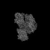 14528MC M: map data used to model this data C: citing same article ( |
|---|---|
| Similar structure data |
-Links
 PDBj
PDBj


- Assembly
| Deposited unit | 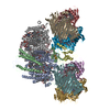
|
|---|---|
| 1 |
|
-Components
-Photosystem P840 reaction ... , 2 types, 3 molecules AaB
| #1: Protein | Mass: 81784.641 Da / Num. of mol.: 2 / Source method: isolated from a natural source / Source: (natural) Chlorobaculum tepidum TLS (bacteria) / Strain: ATCC 49652 / DSM 12025 / NBRC 103806 / TLS / References: UniProt: Q8KAY0#2: Protein | | Mass: 23540.289 Da / Num. of mol.: 1 / Source method: isolated from a natural source / Source: (natural) Chlorobaculum tepidum TLS (bacteria) / Strain: ATCC 49652 / DSM 12025 / NBRC 103806 / TLS / References: UniProt: Q8KAY1 |
|---|
-Protein , 2 types, 3 molecules CcD
| #3: Protein | Mass: 22741.779 Da / Num. of mol.: 2 / Source method: isolated from a natural source / Source: (natural) Chlorobaculum tepidum TLS (bacteria) / Strain: ATCC 49652 / DSM 12025 / NBRC 103806 / TLS / References: UniProt: O07091#4: Protein | | Mass: 16633.195 Da / Num. of mol.: 1 / Source method: isolated from a natural source / Source: (natural) Chlorobaculum tepidum TLS (bacteria) / Strain: ATCC 49652 / DSM 12025 / NBRC 103806 / TLS / References: UniProt: Q8KEP5 |
|---|
-Bacteriochlorophyll ... , 1 types, 6 molecules EFGHIJ
| #5: Protein | Mass: 40343.430 Da / Num. of mol.: 6 / Source method: isolated from a natural source / Source: (natural) Chlorobaculum tepidum TLS (bacteria) / Strain: ATCC 49652 / DSM 12025 / NBRC 103806 / TLS / References: UniProt: Q46393 |
|---|
-Non-polymers , 9 types, 101 molecules 


| #6: Chemical |  Mass: 911.504 Da / Num. of mol.: 2 / Source method: obtained synthetically / Formula: C55H74MgN4O6 / Feature type: SUBJECT OF INVESTIGATION Mass: 911.504 Da / Num. of mol.: 2 / Source method: obtained synthetically / Formula: C55H74MgN4O6 / Feature type: SUBJECT OF INVESTIGATION#7: Chemical | ChemComp-CLA /  Mass: 893.489 Da / Num. of mol.: 4 / Source method: obtained synthetically / Formula: C55H72MgN4O5 / Feature type: SUBJECT OF INVESTIGATION Mass: 893.489 Da / Num. of mol.: 4 / Source method: obtained synthetically / Formula: C55H72MgN4O5 / Feature type: SUBJECT OF INVESTIGATION#8: Chemical | ChemComp-BCL /  Mass: 911.504 Da / Num. of mol.: 72 / Source method: obtained synthetically / Formula: C55H74MgN4O6 / Feature type: SUBJECT OF INVESTIGATION Mass: 911.504 Da / Num. of mol.: 72 / Source method: obtained synthetically / Formula: C55H74MgN4O6 / Feature type: SUBJECT OF INVESTIGATION#9: Chemical |  Mass: 895.299 Da / Num. of mol.: 2 / Source method: obtained synthetically / Formula: C58H86O7 / Feature type: SUBJECT OF INVESTIGATION Mass: 895.299 Da / Num. of mol.: 2 / Source method: obtained synthetically / Formula: C58H86O7 / Feature type: SUBJECT OF INVESTIGATION#10: Chemical | ChemComp-LHG /  Mass: 722.970 Da / Num. of mol.: 4 / Source method: obtained synthetically / Formula: C38H75O10P / Feature type: SUBJECT OF INVESTIGATION / Comment: phospholipid*YM Mass: 722.970 Da / Num. of mol.: 4 / Source method: obtained synthetically / Formula: C38H75O10P / Feature type: SUBJECT OF INVESTIGATION / Comment: phospholipid*YM#11: Chemical |  Mass: 731.052 Da / Num. of mol.: 2 / Source method: obtained synthetically / Formula: C41H78O10 / Feature type: SUBJECT OF INVESTIGATION Mass: 731.052 Da / Num. of mol.: 2 / Source method: obtained synthetically / Formula: C41H78O10 / Feature type: SUBJECT OF INVESTIGATION#12: Chemical |  Mass: 40.078 Da / Num. of mol.: 2 / Source method: obtained synthetically / Formula: Ca / Feature type: SUBJECT OF INVESTIGATION Mass: 40.078 Da / Num. of mol.: 2 / Source method: obtained synthetically / Formula: Ca / Feature type: SUBJECT OF INVESTIGATION#13: Chemical |  Mass: 351.640 Da / Num. of mol.: 3 / Source method: obtained synthetically / Formula: Fe4S4 / Feature type: SUBJECT OF INVESTIGATION Mass: 351.640 Da / Num. of mol.: 3 / Source method: obtained synthetically / Formula: Fe4S4 / Feature type: SUBJECT OF INVESTIGATION#14: Water | ChemComp-HOH / | Mass: 18.015 Da / Num. of mol.: 10 / Source method: isolated from a natural source / Formula: H2O |
|---|
-Details
| Has ligand of interest | Y |
|---|
-Experimental details
-Experiment
| Experiment | Method: ELECTRON MICROSCOPY |
|---|---|
| EM experiment | Aggregation state: PARTICLE / 3D reconstruction method: single particle reconstruction |
- Sample preparation
| Component | Name: Photosystem P840 reaction center / Type: COMPLEX / Entity ID: #1-#5 / Source: NATURAL |
|---|---|
| Molecular weight | Value: 0.49 MDa / Experimental value: YES |
| Source (natural) | Organism: Chlorobaculum tepidum TLS (bacteria) |
| Buffer solution | pH: 8 |
| Specimen | Embedding applied: NO / Shadowing applied: NO / Staining applied: NO / Vitrification applied: YES |
| Specimen support | Grid material: COPPER / Grid mesh size: 200 divisions/in. / Grid type: Quantifoil |
| Vitrification | Instrument: FEI VITROBOT MARK IV / Cryogen name: ETHANE / Humidity: 100 % / Chamber temperature: 4 K |
- Electron microscopy imaging
| Experimental equipment |  Model: Titan Krios / Image courtesy: FEI Company |
|---|---|
| Microscopy | Model: FEI TITAN KRIOS |
| Electron gun | Electron source:  FIELD EMISSION GUN / Accelerating voltage: 300 kV / Illumination mode: FLOOD BEAM FIELD EMISSION GUN / Accelerating voltage: 300 kV / Illumination mode: FLOOD BEAM |
| Electron lens | Mode: BRIGHT FIELD / Nominal magnification: 105000 X / Nominal defocus max: 2500 nm / Nominal defocus min: 1200 nm / Calibrated defocus min: 1200 nm / Calibrated defocus max: 2500 nm / Cs: 2.7 mm / Alignment procedure: COMA FREE |
| Specimen holder | Cryogen: NITROGEN / Specimen holder model: FEI TITAN KRIOS AUTOGRID HOLDER |
| Image recording | Electron dose: 45 e/Å2 / Film or detector model: GATAN K3 (6k x 4k) |
| Image scans | Width: 4092 / Height: 5769 |
- Processing
| EM software |
| ||||||||||||||||||||||||
|---|---|---|---|---|---|---|---|---|---|---|---|---|---|---|---|---|---|---|---|---|---|---|---|---|---|
| CTF correction | Type: PHASE FLIPPING AND AMPLITUDE CORRECTION | ||||||||||||||||||||||||
| Particle selection | Num. of particles selected: 4791019 | ||||||||||||||||||||||||
| 3D reconstruction | Resolution: 2.5 Å / Resolution method: FSC 0.143 CUT-OFF / Num. of particles: 481095 / Symmetry type: POINT | ||||||||||||||||||||||||
| Atomic model building | B value: 60 | ||||||||||||||||||||||||
| Atomic model building |
|


