Movie
Movie Controller

[English] 日本語
 Yorodumi
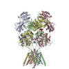
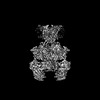
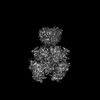
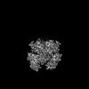
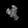
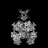
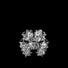
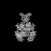
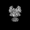
- PDB-7yfl: Structure of GluN1a-GluN2D NMDA receptor in complex with agonists... -
+ Open data

- Basic information
| Entry | Database: PDB / ID: 7yfl | ||||||||||||||||||
|---|---|---|---|---|---|---|---|---|---|---|---|---|---|---|---|---|---|---|---|
| Title | Structure of GluN1a-GluN2D NMDA receptor in complex with agonists glycine and glutamate. | ||||||||||||||||||
 Components Components | (Glutamate receptor ionotropic, NMDA ...) x 2 | ||||||||||||||||||
 Keywords Keywords | ELECTRON TRANSPORT / ion channel / cryo-EM structure / glutamate receptor / synaptic protein | ||||||||||||||||||
| Function / homology |  Function and homology information Function and homology informationglycine-gated cation channel activity / regulation of sensory perception of pain / excitatory chemical synaptic transmission / Synaptic adhesion-like molecules / cellular response to L-glutamate / propylene metabolic process / response to glycine / Assembly and cell surface presentation of NMDA receptors / neurotransmitter receptor complex / Neurexins and neuroligins ...glycine-gated cation channel activity / regulation of sensory perception of pain / excitatory chemical synaptic transmission / Synaptic adhesion-like molecules / cellular response to L-glutamate / propylene metabolic process / response to glycine / Assembly and cell surface presentation of NMDA receptors / neurotransmitter receptor complex / Neurexins and neuroligins / regulation of monoatomic cation transmembrane transport / NMDA glutamate receptor activity / NMDA selective glutamate receptor complex / glutamate binding / voltage-gated monoatomic cation channel activity / ligand-gated sodium channel activity / calcium ion transmembrane import into cytosol / protein heterotetramerization / glycine binding / startle response / positive regulation of reactive oxygen species biosynthetic process / Negative regulation of NMDA receptor-mediated neuronal transmission / Unblocking of NMDA receptors, glutamate binding and activation / Long-term potentiation / monoatomic cation transmembrane transport / positive regulation of calcium ion transport into cytosol / regulation of neuronal synaptic plasticity / monoatomic cation transport / ligand-gated monoatomic ion channel activity / synaptic cleft / calcium ion homeostasis / glutamate-gated receptor activity / positive regulation of synaptic transmission, glutamatergic / EPHB-mediated forward signaling / glutamate-gated calcium ion channel activity / presynaptic active zone membrane / excitatory synapse / ionotropic glutamate receptor signaling pathway / ligand-gated monoatomic ion channel activity involved in regulation of presynaptic membrane potential / Ras activation upon Ca2+ influx through NMDA receptor / positive regulation of excitatory postsynaptic potential / hippocampal mossy fiber to CA3 synapse / sodium ion transmembrane transport / synaptic membrane / adult locomotory behavior / transmitter-gated monoatomic ion channel activity involved in regulation of postsynaptic membrane potential / synaptic transmission, glutamatergic / regulation of membrane potential / excitatory postsynaptic potential / brain development / visual learning / regulation of synaptic plasticity / postsynaptic density membrane / calcium ion transmembrane transport / terminal bouton / synaptic vesicle / long-term synaptic potentiation / amyloid-beta binding / signaling receptor activity / RAF/MAP kinase cascade / dendritic spine / chemical synaptic transmission / response to ethanol / calmodulin binding / postsynaptic membrane / neuron projection / postsynaptic density / calcium ion binding / synapse / dendrite / endoplasmic reticulum membrane / protein-containing complex binding / glutamatergic synapse / cell surface / positive regulation of transcription by RNA polymerase II / plasma membrane / cytoplasm Similarity search - Function | ||||||||||||||||||
| Biological species |  Homo sapiens (human) Homo sapiens (human) | ||||||||||||||||||
| Method | ELECTRON MICROSCOPY / single particle reconstruction / cryo EM / Resolution: 3.9 Å | ||||||||||||||||||
 Authors Authors | Zhang, J.L. / Zhu, S.J. / Zhang, M. | ||||||||||||||||||
| Funding support |  China, 5items China, 5items
| ||||||||||||||||||
 Citation Citation | Journal: Nat Struct Mol Biol / Year: 2023 Title: Distinct structure and gating mechanism in diverse NMDA receptors with GluN2C and GluN2D subunits. Authors: Jilin Zhang / Ming Zhang / Qinrui Wang / Han Wen / Zheyi Liu / Fangjun Wang / Yuhang Wang / Fenyong Yao / Nan Song / Zengwei Kou / Yang Li / Fei Guo / Shujia Zhu / Abstract: N-methyl-D-aspartate (NMDA) receptors are heterotetramers comprising two GluN1 and two alternate GluN2 (N2A-N2D) subunits. Here we report full-length cryo-EM structures of the human N1-N2D di- ...N-methyl-D-aspartate (NMDA) receptors are heterotetramers comprising two GluN1 and two alternate GluN2 (N2A-N2D) subunits. Here we report full-length cryo-EM structures of the human N1-N2D di-heterotetramer (di-receptor), rat N1-N2C di-receptor and N1-N2A-N2C tri-heterotetramer (tri-receptor) at a best resolution of 3.0 Å. The bilobate N-terminal domain (NTD) in N2D intrinsically adopts a closed conformation, leading to a compact NTD tetramer in the N1-N2D receptor. Additionally, crosslinking the ligand-binding domain (LBD) of two N1 protomers significantly elevated the channel open probability (Po) in N1-N2D di-receptors. Surprisingly, the N1-N2C di-receptor adopted both symmetric (minor) and asymmetric (major) conformations, the latter further locked by an allosteric potentiator, PYD-106, binding to a pocket between the NTD and LBD in only one N2C protomer. Finally, the N2A and N2C subunits in the N1-N2A-N2C tri-receptor display a conformation close to one protomer in the N1-N2A and N1-N2C di-receptors, respectively. These findings provide a comprehensive structural understanding of diverse function in major NMDA receptor subtypes. | ||||||||||||||||||
| History |
|
- Structure visualization
| Structure viewer | Molecule: MolmilJmol/JSmol |
|---|
- Downloads & links
-Download
| PDBx/mmCIF format | 7yfl.cif.gz | 477.9 KB | Display | PDBx/mmCIF format |
|---|---|---|---|---|
| PDB format | pdb7yfl.ent.gz | 370.8 KB | Display | PDB format |
| PDBx/mmJSON format | 7yfl.json.gz | Tree view | PDBx/mmJSON format | |
| Others |  Other downloads Other downloads |
-Validation report
| Arichive directory | https://data.pdbj.org/pub/pdb/validation_reports/yf/7yflftp://data.pdbj.org/pub/pdb/validation_reports/yf/7yfl | HTTPS FTP |
|---|
-Related structure data
| Related structure data | 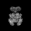 33792MC  7yffC  7yfgC  7yfhC  7yfiC  7yfmC  7yfoC  7yfrC  8hdkC C: citing same article ( M: map data used to model this data |
|---|---|
| Similar structure data |
-Links
 PDBj
PDBj


- Assembly
| Deposited unit | 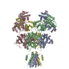
|
|---|---|
| 1 |
|
-Components
-Glutamate receptor ionotropic, NMDA ... , 2 types, 4 molecules ACBD
| #1: Protein | Mass: 95236.078 Da / Num. of mol.: 2 Source method: isolated from a genetically manipulated source Source: (gene. exp.) Homo sapiens (human) / Gene: GRIN1, NMDAR1 / Plasmid: pEG-BacMam / Cell (production host): Kidney embryonic cells / Cell line (production host): HEK293S GnTI- / Production host: Homo sapiens (human) / References: UniProt: Q05586#2: Protein | Mass: 97226.414 Da / Num. of mol.: 2 Source method: isolated from a genetically manipulated source Source: (gene. exp.) Homo sapiens (human) / Gene: GRIN2D, GluN2D, NMDAR2D / Plasmid: pEG-BacMam / Cell (production host): Kidney embryonic cells / Cell line (production host): HEK293S GnTI- / Production host: Homo sapiens (human) / References: UniProt: O15399 |
|---|
-Sugars , 2 types, 14 molecules 
| #3: Polysaccharide | 2-acetamido-2-deoxy-beta-D-glucopyranose-(1-4)-2-acetamido-2-deoxy-beta-D-glucopyranose Source method: isolated from a genetically manipulated source #5: Sugar | ChemComp-NAG /  Type: D-saccharide, beta linking / Mass: 221.208 Da / Num. of mol.: 10 / Source method: obtained synthetically / Formula: C8H15NO6 / Feature type: SUBJECT OF INVESTIGATION Type: D-saccharide, beta linking / Mass: 221.208 Da / Num. of mol.: 10 / Source method: obtained synthetically / Formula: C8H15NO6 / Feature type: SUBJECT OF INVESTIGATION |
|---|
-Non-polymers , 2 types, 4 molecules 

| #4: Chemical |  Type: peptide linking / Mass: 75.067 Da / Num. of mol.: 2 / Source method: obtained synthetically / Formula: C2H5NO2 Type: peptide linking / Mass: 75.067 Da / Num. of mol.: 2 / Source method: obtained synthetically / Formula: C2H5NO2#6: Chemical |  Type: L-peptide linking / Mass: 147.129 Da / Num. of mol.: 2 / Source method: obtained synthetically / Formula: C5H9NO4 Type: L-peptide linking / Mass: 147.129 Da / Num. of mol.: 2 / Source method: obtained synthetically / Formula: C5H9NO4 |
|---|
-Details
| Has ligand of interest | Y |
|---|---|
| Has protein modification | Y |
-Experimental details
-Experiment
| Experiment | Method: ELECTRON MICROSCOPY |
|---|---|
| EM experiment | Aggregation state: PARTICLE / 3D reconstruction method: single particle reconstruction |
- Sample preparation
| Component | Name: NMDA receptor with NMDA 1 incorperated with NMDA 2D / Type: COMPLEX / Entity ID: #1-#2 / Source: RECOMBINANT |
|---|---|
| Molecular weight | Value: 384.54 kDa/nm / Experimental value: NO |
| Source (natural) | Organism: Homo sapiens (human) / Strain: Homo sapiens / Cellular location: plasma membrane / Organ: brain / Organelle: synapse / Tissue: brain |
| Source (recombinant) | Organism: Homo sapiens (human) / Strain: Homo sapiens / Cell: Kidney embryonic cells / Plasmid: pEG-BacMam |
| Buffer solution | pH: 8 |
| Specimen | Conc.: 4 mg/ml / Embedding applied: NO / Shadowing applied: NO / Staining applied: NO / Vitrification applied: YES / Details: This sample was monodisperse |
| Specimen support | Grid material: GOLD / Grid mesh size: 300 divisions/in. / Grid type: Quantifoil R1.2/1.3 |
| Vitrification | Instrument: FEI VITROBOT MARK IV / Cryogen name: ETHANE / Humidity: 100 % / Chamber temperature: 281 K / Details: Blot for 3 seconds before plunging |
- Electron microscopy imaging
| Experimental equipment |  Model: Titan Krios / Image courtesy: FEI Company |
|---|---|
| Microscopy | Model: FEI TITAN KRIOS |
| Electron gun | Electron source:  FIELD EMISSION GUN / Accelerating voltage: 300 kV / Illumination mode: FLOOD BEAM FIELD EMISSION GUN / Accelerating voltage: 300 kV / Illumination mode: FLOOD BEAM |
| Electron lens | Mode: BRIGHT FIELD / Nominal defocus max: 2500 nm / Nominal defocus min: 1500 nm |
| Image recording | Electron dose: 60 e/Å2 / Detector mode: SUPER-RESOLUTION / Film or detector model: DIRECT ELECTRON DE-10 (5k x 4k) / Num. of grids imaged: 1 / Num. of real images: 3840 |
| Image scans | Movie frames/image: 40 / Used frames/image: 1-40 |
- Processing
| Software | Name: PHENIX / Version: 1.20.1_4487: / Classification: refinement | ||||||||||||||||||||||||||||||||||||
|---|---|---|---|---|---|---|---|---|---|---|---|---|---|---|---|---|---|---|---|---|---|---|---|---|---|---|---|---|---|---|---|---|---|---|---|---|---|
| EM software |
| ||||||||||||||||||||||||||||||||||||
| CTF correction | Type: NONE | ||||||||||||||||||||||||||||||||||||
| Particle selection | Num. of particles selected: 505204 | ||||||||||||||||||||||||||||||||||||
| Symmetry | Point symmetry: C2 (2 fold cyclic) | ||||||||||||||||||||||||||||||||||||
| 3D reconstruction | Resolution: 3.9 Å / Resolution method: FSC 0.143 CUT-OFF / Num. of particles: 232194 / Num. of class averages: 2 / Symmetry type: POINT | ||||||||||||||||||||||||||||||||||||
| Atomic model building | B value: 220 / Protocol: FLEXIBLE FIT / Space: REAL / Target criteria: Correlation coefficient | ||||||||||||||||||||||||||||||||||||
| Atomic model building | PDB-ID: 6WI1 Accession code: 6WI1 / Source name: PDB / Type: experimental model | ||||||||||||||||||||||||||||||||||||
| Refine LS restraints |
|