 Movie
Movie Controller


+ Open data

- Basic information
| Entry | Database: PDB / ID: 7y1a | ||||||
|---|---|---|---|---|---|---|---|
| Title | Lateral hexamer | ||||||
 Components Components |
| ||||||
 Keywords Keywords | PHOTOSYNTHESIS / Lateral hexamer / PBS | ||||||
| Function / homology |  Function and homology information Function and homology information | ||||||
| Biological species |  Porphyridium purpureum (eukaryote) Porphyridium purpureum (eukaryote) | ||||||
| Method | ELECTRON MICROSCOPY / single particle reconstruction / cryo EM / Resolution: 6.3 Å | ||||||
 Authors Authors | You, X. / Zhang, X. / Cheng, J. / Xiao, Y.N. / Sun, S. / Sui, S.F. | ||||||
| Funding support | 1items
| ||||||
 Citation Citation | Journal: Nature / Year: 2023 Title: In situ structure of the red algal phycobilisome-PSII-PSI-LHC megacomplex. Authors: Xin You / Xing Zhang / Jing Cheng / Yanan Xiao / Jianfei Ma / Shan Sun / Xinzheng Zhang / Hong-Wei Wang / Sen-Fang Sui /  Abstract: In oxygenic photosynthetic organisms, light energy is captured by antenna systems and transferred to photosystem II (PSII) and photosystem I (PSI) to drive photosynthesis. The antenna systems of red ...In oxygenic photosynthetic organisms, light energy is captured by antenna systems and transferred to photosystem II (PSII) and photosystem I (PSI) to drive photosynthesis. The antenna systems of red algae consist of soluble phycobilisomes (PBSs) and transmembrane light-harvesting complexes (LHCs). Excitation energy transfer pathways from PBS to photosystems remain unclear owing to the lack of structural information. Here we present in situ structures of PBS-PSII-PSI-LHC megacomplexes from the red alga Porphyridium purpureum at near-atomic resolution using cryogenic electron tomography and in situ single-particle analysis, providing interaction details between PBS, PSII and PSI. The structures reveal several unidentified and incomplete proteins and their roles in the assembly of the megacomplex, as well as a huge and sophisticated pigment network. This work provides a solid structural basis for unravelling the mechanisms of PBS-PSII-PSI-LHC megacomplex assembly, efficient energy transfer from PBS to the two photosystems, and regulation of energy distribution between PSII and PSI. | ||||||
| History |
|
- Structure visualization
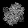
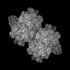
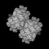
| Structure viewer | Molecule: MolmilJmol/JSmol |
|---|
- Downloads & links
-Download
| PDBx/mmCIF format | 7y1a.cif.gz | 381.7 KB | Display | PDBx/mmCIF format |
|---|---|---|---|---|
| PDB format | pdb7y1a.ent.gz | 332.2 KB | Display | PDB format |
| PDBx/mmJSON format | 7y1a.json.gz | Tree view | PDBx/mmJSON format | |
| Others |  Other downloads Other downloads |
-Validation report
| Arichive directory | https://data.pdbj.org/pub/pdb/validation_reports/y1/7y1aftp://data.pdbj.org/pub/pdb/validation_reports/y1/7y1a | HTTPS FTP |
|---|
-Related structure data
| Related structure data |  33558MC  7y4lC  7y5eC 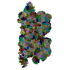 7y7aC M: map data used to model this data C: citing same article ( |
|---|---|
| Similar structure data |

-Links
 PDBj
PDBj- Assembly
| Deposited unit | 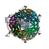
|
|---|---|
| 1 |
|
-Components
| #1: Protein | Mass: 18584.182 Da / Num. of mol.: 6 Source method: isolated from a genetically manipulated source Source: (gene. exp.) Porphyridium purpureum (eukaryote) / Gene: cpeB / Production host: Porphyridium purpureum (eukaryote) / References: UniProt: P11393#2: Protein | Mass: 17824.029 Da / Num. of mol.: 6 Source method: isolated from a genetically manipulated source Source: (gene. exp.) Porphyridium purpureum (eukaryote) / Gene: peA, cpeA / Production host: Porphyridium purpureum (eukaryote) / References: UniProt: E2IH77#3: Protein | | Mass: 7422.140 Da / Num. of mol.: 1 Source method: isolated from a genetically manipulated source Source: (gene. exp.) Porphyridium purpureum (eukaryote) / Production host: Porphyridium purpureum (eukaryote)#4: Protein | | Mass: 12783.750 Da / Num. of mol.: 1 Source method: isolated from a genetically manipulated source Source: (gene. exp.) Porphyridium purpureum (eukaryote) / Production host: Porphyridium purpureum (eukaryote)#5: Chemical | ChemComp-PEB /   Mass: 588.694 Da / Num. of mol.: 31 / Source method: obtained synthetically / Formula: C33H40N4O6 / Feature type: SUBJECT OF INVESTIGATION Mass: 588.694 Da / Num. of mol.: 31 / Source method: obtained synthetically / Formula: C33H40N4O6 / Feature type: SUBJECT OF INVESTIGATIONHas ligand of interest | Y | |
|---|
-Experimental details
-Experiment
| Experiment | Method: ELECTRON MICROSCOPY |
|---|---|
| EM experiment | Aggregation state: CELL / 3D reconstruction method: single particle reconstruction |
- Sample preparation
| Component | Name: Lateral hexamer of PBS / Type: COMPLEX / Entity ID: #1-#4 / Source: NATURAL |
|---|---|
| Source (natural) | Organism: Porphyridium purpureum (eukaryote) |
| Buffer solution | pH: 7 |
| Specimen | Embedding applied: NO / Shadowing applied: NO / Staining applied: NO / Vitrification applied: YES |
| Vitrification | Cryogen name: ETHANE |
- Electron microscopy imaging
| Experimental equipment |  Model: Titan Krios / Image courtesy: FEI Company |
|---|---|
| Microscopy | Model: FEI TITAN KRIOS |
| Electron gun | Electron source:  FIELD EMISSION GUN / Accelerating voltage: 300 kV / Illumination mode: SPOT SCAN FIELD EMISSION GUN / Accelerating voltage: 300 kV / Illumination mode: SPOT SCAN |
| Electron lens | Mode: BRIGHT FIELD / Nominal defocus max: 6000 nm / Nominal defocus min: 1000 nm |
| Image recording | Electron dose: 35 e/Å2 / Film or detector model: GATAN K3 (6k x 4k) |
- Processing
| CTF correction | Type: PHASE FLIPPING AND AMPLITUDE CORRECTION |
|---|---|
| 3D reconstruction | Resolution: 6.3 Å / Resolution method: FSC 0.143 CUT-OFF / Num. of particles: 87000 / Symmetry type: POINT |
