 Movie
Movie Controller

[English] 日本語
 Yorodumi
- PDB-7xbw: Cryo-EM structure of the human chemokine receptor CX3CR1 in compl... -
+ Open data

- Basic information
| Entry | Database: PDB / ID: 7xbw | ||||||||||||
|---|---|---|---|---|---|---|---|---|---|---|---|---|---|
| Title | Cryo-EM structure of the human chemokine receptor CX3CR1 in complex with Gi1 | ||||||||||||
 Components Components |
| ||||||||||||
 Keywords Keywords | SIGNALING PROTEIN / G protein-coupled receptor / chemokine receptor / CX3CR1 | ||||||||||||
| Function / homology |  Function and homology information Function and homology informationC-X3-C chemokine receptor activity / dendritic tree / multiple spine synapse organization, single dendrite / negative regulation of microglial cell mediated cytotoxicity / macropinosome membrane / C-X3-C chemokine binding / regulation of microglial cell migration / CX3C chemokine receptor binding / autocrine signaling / host-mediated modulation of intestinal microbiota composition ...C-X3-C chemokine receptor activity / dendritic tree / multiple spine synapse organization, single dendrite / negative regulation of microglial cell mediated cytotoxicity / macropinosome membrane / C-X3-C chemokine binding / regulation of microglial cell migration / CX3C chemokine receptor binding / autocrine signaling / host-mediated modulation of intestinal microbiota composition / microglial cell activation involved in immune response / synapse pruning / central nervous system maturation / synapse maturation / negative regulation of hippocampal neuron apoptotic process / antifungal innate immune response / chemokine receptor activity / leukocyte tethering or rolling / regulation of tumor necrosis factor production / C-C chemokine receptor activity / C-C chemokine binding / positive regulation of monocyte chemotaxis / G protein-coupled peptide receptor activity / leukocyte chemotaxis / regulation of nitric oxide biosynthetic process / Chemokine receptors bind chemokines / negative regulation of interleukin-1 beta production / positive regulation of neurogenesis / positive regulation of neuroblast proliferation / RSV-host interactions / neuronal cell body membrane / Respiratory syncytial virus (RSV) attachment and entry / negative regulation of apoptotic signaling pathway / regulation of neurogenesis / social behavior / cellular defense response / adenylate cyclase inhibitor activity / positive regulation of protein localization to cell cortex / T cell migration / positive regulation of relaxation of smooth muscle / Adenylate cyclase inhibitory pathway / D2 dopamine receptor binding / adenylate cyclase-inhibiting serotonin receptor signaling pathway / G protein-coupled serotonin receptor binding / negative regulation of angiogenesis / response to ischemia / cellular response to forskolin / regulation of mitotic spindle organization / chemokine-mediated signaling pathway / cell chemotaxis / Regulation of insulin secretion / calcium-mediated signaling / neuropeptide signaling pathway / response to prostaglandin E / brain development / positive regulation of cholesterol biosynthetic process / negative regulation of insulin secretion / response to wounding / regulation of synaptic plasticity / G protein-coupled receptor binding / modulation of chemical synaptic transmission / response to peptide hormone / G protein-coupled receptor activity / chemotaxis / centriolar satellite / G-protein beta/gamma-subunit complex binding / adenylate cyclase-modulating G protein-coupled receptor signaling pathway / adenylate cyclase-inhibiting G protein-coupled receptor signaling pathway / Olfactory Signaling Pathway / Activation of the phototransduction cascade / G protein-coupled acetylcholine receptor signaling pathway / G beta:gamma signalling through PLC beta / Presynaptic function of Kainate receptors / Thromboxane signalling through TP receptor / Activation of G protein gated Potassium channels / Inhibition of voltage gated Ca2+ channels via Gbeta/gamma subunits / G-protein activation / Glucagon signaling in metabolic regulation / Prostacyclin signalling through prostacyclin receptor / G beta:gamma signalling through CDC42 / Synthesis, secretion, and inactivation of Glucagon-like Peptide-1 (GLP-1) / G beta:gamma signalling through BTK / photoreceptor disc membrane / ADP signalling through P2Y purinoceptor 12 / Glucagon-type ligand receptors / Sensory perception of sweet, bitter, and umami (glutamate) taste / GDP binding / Adrenaline,noradrenaline inhibits insulin secretion / Vasopressin regulates renal water homeostasis via Aquaporins / Glucagon-like Peptide-1 (GLP1) regulates insulin secretion / G alpha (z) signalling events / cellular response to catecholamine stimulus / ADP signalling through P2Y purinoceptor 1 / ADORA2B mediated anti-inflammatory cytokines production / G beta:gamma signalling through PI3Kgamma / cell-cell signaling / adenylate cyclase-activating dopamine receptor signaling pathway / Cooperation of PDCL (PhLP1) and TRiC/CCT in G-protein beta folding / GPER1 signaling / cellular response to prostaglandin E stimulus Similarity search - Function | ||||||||||||
| Biological species |  Homo sapiens (human) Homo sapiens (human) | ||||||||||||
| Method | ELECTRON MICROSCOPY / single particle reconstruction / cryo EM / Resolution: 2.8 Å | ||||||||||||
 Authors Authors | Lu, M. / Zhao, W. / Han, S. / Zhu, Y. / Wu, B. / Zhao, Q. | ||||||||||||
| Funding support |  China, 3items China, 3items
| ||||||||||||
 Citation Citation | Journal: Sci Adv / Year: 2022 Title: Activation of the human chemokine receptor CX3CR1 regulated by cholesterol. Authors: Minmin Lu / Wenli Zhao / Shuo Han / Xiaowen Lin / Tingyu Xu / Qiuxiang Tan / Mu Wang / Cuiying Yi / Xiaojing Chu / Weibo Yang / Ya Zhu / Beili Wu / Qiang Zhao / Abstract: As the only member of the CX3C chemokine receptor subfamily, CX3CR1 binds to its sole endogenous ligand CX3CL1, which shows notable potential as a therapeutic target in atherosclerosis, cancer, and ...As the only member of the CX3C chemokine receptor subfamily, CX3CR1 binds to its sole endogenous ligand CX3CL1, which shows notable potential as a therapeutic target in atherosclerosis, cancer, and neuropathy. However, the drug development of CX3CR1 is hampered partially by the lack of structural information. Here, we present two cryo-electron microscopy structures of CX3CR1-G complexes in ligand-free and CX3CL1-bound states at 2.8- and 3.4-Å resolution, respectively. Together with functional data, the structures reveal the key factors that govern the recognition of CX3CL1 by both CX3CR1 and US28. A much smaller conformational change of helix VI upon activation than previously solved class A GPCR-G complex structures is observed in CX3CR1, which may correlate with three cholesterol molecules that play essential roles in conformation stabilization and signaling transduction. Thus, our data deepen the understanding of cholesterol modulation in GPCR (G protein-coupled receptor) signaling and provide insights into the diversity of G protein coupling. | ||||||||||||
| History |
|
- Structure visualization
| Structure viewer | Molecule: MolmilJmol/JSmol |
|---|
- Downloads & links
-Download
| PDBx/mmCIF format | 7xbw.cif.gz | 174.2 KB | Display | PDBx/mmCIF format |
|---|---|---|---|---|
| PDB format | pdb7xbw.ent.gz | 126.1 KB | Display | PDB format |
| PDBx/mmJSON format | 7xbw.json.gz | Tree view | PDBx/mmJSON format | |
| Others |  Other downloads Other downloads |
-Validation report
| Arichive directory | https://data.pdbj.org/pub/pdb/validation_reports/xb/7xbwftp://data.pdbj.org/pub/pdb/validation_reports/xb/7xbw | HTTPS FTP |
|---|
-Related structure data
| Related structure data |  33107MC  7xbxC M: map data used to model this data C: citing same article ( |
|---|---|
| Similar structure data |
-Links
 PDBj
PDBj


- Assembly
| Deposited unit | 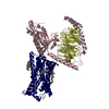
|
|---|---|
| 1 |
|
-Components
| #1: Protein | Mass: 40447.141 Da / Num. of mol.: 1 / Mutation: S47C,G202T,G203A,E245A,A326S Source method: isolated from a genetically manipulated source Source: (gene. exp.) Homo sapiens (human) / Gene: GNAI1 / Production host:   Spodoptera frugiperda (fall armyworm) / References: UniProt: P63096 Spodoptera frugiperda (fall armyworm) / References: UniProt: P63096 | ||||
|---|---|---|---|---|---|
| #2: Protein | Mass: 38245.805 Da / Num. of mol.: 1 Source method: isolated from a genetically manipulated source Source: (gene. exp.) Homo sapiens (human) / Gene: GNB1 / Production host: Spodoptera frugiperda (fall armyworm) / References: UniProt: P62873 | ||||
| #3: Protein | Mass: 7861.143 Da / Num. of mol.: 1 Source method: isolated from a genetically manipulated source Source: (gene. exp.) Homo sapiens (human) / Gene: GNG2 / Production host: Spodoptera frugiperda (fall armyworm) / References: UniProt: P59768 | ||||
| #4: Protein | Mass: 41072.680 Da / Num. of mol.: 1 / Mutation: I120L, C221S, M250V Source method: isolated from a genetically manipulated source Source: (gene. exp.) Homo sapiens (human) / Gene: CX3CR1, CMKBRL1, GPR13 / Production host: Spodoptera frugiperda (fall armyworm) / References: UniProt: P49238 | ||||
| #5: Chemical |   Mass: 386.654 Da / Num. of mol.: 3 / Source method: obtained synthetically / Formula: C27H46O Mass: 386.654 Da / Num. of mol.: 3 / Source method: obtained synthetically / Formula: C27H46OHas ligand of interest | N | Has protein modification | Y | |
-Experimental details
-Experiment
| Experiment | Method: ELECTRON MICROSCOPY |
|---|---|
| EM experiment | Aggregation state: PARTICLE / 3D reconstruction method: single particle reconstruction |
- Sample preparation
| Component | Name: Chemokine receptor CX3CR1 in complex with Gi1 / Type: COMPLEX / Entity ID: #1-#4 / Source: RECOMBINANT |
|---|---|
| Source (natural) | Organism: Homo sapiens (human) |
| Source (recombinant) | Organism: Spodoptera frugiperda (fall armyworm) |
| Buffer solution | pH: 7.5 |
| Specimen | Embedding applied: NO / Shadowing applied: NO / Staining applied: NO / Vitrification applied: YES |
| Vitrification | Cryogen name: ETHANE |
- Electron microscopy imaging
| Experimental equipment |  Model: Titan Krios / Image courtesy: FEI Company |
|---|---|
| Microscopy | Model: FEI TITAN KRIOS |
| Electron gun | Electron source:  FIELD EMISSION GUN / Accelerating voltage: 300 kV / Illumination mode: SPOT SCAN FIELD EMISSION GUN / Accelerating voltage: 300 kV / Illumination mode: SPOT SCAN |
| Electron lens | Mode: BRIGHT FIELD / Nominal defocus max: 1500 nm / Nominal defocus min: 800 nm |
| Image recording | Electron dose: 2.1875 e/Å2 / Film or detector model: GATAN K3 BIOQUANTUM (6k x 4k) |
- Processing
| Software |
| ||||||||||||||||||||||||
|---|---|---|---|---|---|---|---|---|---|---|---|---|---|---|---|---|---|---|---|---|---|---|---|---|---|
| CTF correction | Type: NONE | ||||||||||||||||||||||||
| 3D reconstruction | Resolution: 2.8 Å / Resolution method: FSC 0.143 CUT-OFF / Num. of particles: 702722 / Symmetry type: POINT | ||||||||||||||||||||||||
| Refinement | Cross valid method: NONE Stereochemistry target values: GeoStd + Monomer Library + CDL v1.2 | ||||||||||||||||||||||||
| Displacement parameters | Biso mean: 52.17 Å2 | ||||||||||||||||||||||||
| Refine LS restraints |
|
