 Movie
Movie Controller

[English] 日本語
 Yorodumi
- PDB-7wqt: Cryo-EM structure of VWF D'D3 dimer complexed with D1D2 at 4.3 an... -
+ Open data

- Basic information
| Entry | Database: PDB / ID: 7wqt | ||||||
|---|---|---|---|---|---|---|---|
| Title | Cryo-EM structure of VWF D'D3 dimer complexed with D1D2 at 4.3 angstron resolution (VWF tube) | ||||||
 Components Components |
| ||||||
 Keywords Keywords | BLOOD CLOTTING / blood / VWF / von Willebrand factor / von Willebrand disease / blood coagulation / multimer assembly / VWF assembly / D'D3 domain / D1D2 domain / D'D3 dimer / D1D2 Dimer / VWF Tube / repeating unit | ||||||
| Function / homology |  Function and homology information Function and homology informationDefective VWF binding to collagen type I / Enhanced cleavage of VWF variant by ADAMTS13 / Defective VWF cleavage by ADAMTS13 variant / Defective F8 binding to von Willebrand factor / hemostasis / Enhanced binding of GP1BA variant to VWF multimer:collagen / Defective binding of VWF variant to GPIb:IX:V / Weibel-Palade body / platelet alpha granule / Platelet Adhesion to exposed collagen ...Defective VWF binding to collagen type I / Enhanced cleavage of VWF variant by ADAMTS13 / Defective VWF cleavage by ADAMTS13 variant / Defective F8 binding to von Willebrand factor / hemostasis / Enhanced binding of GP1BA variant to VWF multimer:collagen / Defective binding of VWF variant to GPIb:IX:V / Weibel-Palade body / platelet alpha granule / Platelet Adhesion to exposed collagen / positive regulation of intracellular signal transduction / GP1b-IX-V activation signalling / p130Cas linkage to MAPK signaling for integrins / cell-substrate adhesion / Defective F8 cleavage by thrombin / Platelet Aggregation (Plug Formation) / immunoglobulin binding / GRB2:SOS provides linkage to MAPK signaling for Integrins / Integrin cell surface interactions / collagen binding / Intrinsic Pathway of Fibrin Clot Formation / Integrin signaling / extracellular matrix / platelet alpha granule lumen / Signaling by high-kinase activity BRAF mutants / MAP2K and MAPK activation / platelet activation / response to wounding / Signaling by RAF1 mutants / Signaling by moderate kinase activity BRAF mutants / Paradoxical activation of RAF signaling by kinase inactive BRAF / Signaling downstream of RAS mutants / Signaling by BRAF and RAF1 fusions / blood coagulation / integrin binding / Platelet degranulation / protein-folding chaperone binding / protease binding / collagen-containing extracellular matrix / cell adhesion / endoplasmic reticulum / extracellular space / extracellular exosome / extracellular region / identical protein binding Similarity search - Function | ||||||
| Biological species |  Homo sapiens (human) Homo sapiens (human) | ||||||
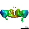
| Method | ELECTRON MICROSCOPY / single particle reconstruction / cryo EM / Resolution: 4.3 Å | ||||||
 Authors Authors | Zeng, J.W. / Shu, Z.M. / Zhou, A.W. | ||||||
| Funding support |  China, 1items China, 1items
| ||||||
 Citation Citation | Journal: Blood / Year: 2022 Title: Structural basis of von Willebrand factor multimerization and tubular storage. Authors: Jianwei Zeng / Zimei Shu / Qian Liang / Jing Zhang / Wenman Wu / Xuefeng Wang / Aiwu Zhou / Abstract: The von Willebrand factor (VWF) propeptide (domains D1D2) is essential for the assembly of VWF multimers and its tubular storage in Weibel-Palade bodies. However, detailed molecular mechanism ...The von Willebrand factor (VWF) propeptide (domains D1D2) is essential for the assembly of VWF multimers and its tubular storage in Weibel-Palade bodies. However, detailed molecular mechanism underlying this propeptide dependence is unclear. Here, we prepared Weibel-Palade body-like tubules using the N-terminal fragment of VWF and solved the cryo-electron microscopy structures of the tubule at atomic resolution. Detailed structural and biochemical analysis indicate that the propeptide forms a homodimer at acidic pH through the D2:D2 binding interface and then recruits 2 D'D3 domains, forming an intertwined D1D2D'D3 homodimer in essence. Stacking of these homodimers by the intermolecular D1:D2 interfaces brings 2 D3 domains face-to-face and facilitates their disulfide linkages and multimerization of VWF. Sequential stacking of these homodimers leads to a right-hand helical tubule for VWF storage. The clinically identified VWF mutations in the propeptide disrupted different steps of the assembling process, leading to diminished VWF multimers in von Willebrand diseases (VWD). Overall, these results indicate that the propeptide serves as a pH-sensing template for VWF multimerization and tubular storage. This sheds light on delivering normal propeptide as a template to rectify the defects in multimerization of VWD mutants. | ||||||
| History |
|
- Structure visualization
| Structure viewer | Molecule: MolmilJmol/JSmol |
|---|
- Downloads & links
-Download
| PDBx/mmCIF format | 7wqt.cif.gz | 3.1 MB | Display | PDBx/mmCIF format |
|---|---|---|---|---|
| PDB format | pdb7wqt.ent.gz | Display | PDB format | |
| PDBx/mmJSON format | 7wqt.json.gz | Tree view | PDBx/mmJSON format | |
| Others |  Other downloads Other downloads |
-Validation report
| Summary document | 7wqt_validation.pdf.gz | 1.6 MB | Display | wwPDB validaton report |
|---|---|---|---|---|
| Full document | 7wqt_full_validation.pdf.gz | 1.6 MB | Display | |
| Data in XML | 7wqt_validation.xml.gz | 394 KB | Display | |
| Data in CIF | 7wqt_validation.cif.gz | 651.9 KB | Display | |
| Arichive directory | https://data.pdbj.org/pub/pdb/validation_reports/wq/7wqtftp://data.pdbj.org/pub/pdb/validation_reports/wq/7wqt | HTTPS FTP |
-Related structure data
| Related structure data |  32713MC  7wppC 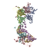 7wpqC  7wprC  7wpsC M: map data used to model this data C: citing same article ( |
|---|---|
| Similar structure data |


-Links
 PDBj
PDBj


- Assembly
| Deposited unit | 
|
|---|---|
| 1 |
|
-Components
| #1: Protein | Mass: 81427.703 Da / Num. of mol.: 16 / Fragment: D1D2 domain Source method: isolated from a genetically manipulated source Source: (gene. exp.) Homo sapiens (human) / Gene: VWF, F8VWF / Production host: Homo sapiens (human) / References: UniProt: P04275#2: Protein | Mass: 54232.809 Da / Num. of mol.: 16 / Fragment: D'D3 domain Source method: isolated from a genetically manipulated source Source: (gene. exp.) Homo sapiens (human) / Gene: VWF, F8VWF / Production host: Homo sapiens (human) / References: UniProt: P04275#3: Sugar | ChemComp-NAG /   Type: D-saccharide, beta linking / Mass: 221.208 Da / Num. of mol.: 42 Type: D-saccharide, beta linking / Mass: 221.208 Da / Num. of mol.: 42Source method: isolated from a genetically manipulated source Formula: C8H15NO6 / Feature type: SUBJECT OF INVESTIGATION #4: Chemical | ChemComp-CA /   Mass: 40.078 Da / Num. of mol.: 56 / Source method: obtained synthetically / Formula: Ca / Feature type: SUBJECT OF INVESTIGATION Mass: 40.078 Da / Num. of mol.: 56 / Source method: obtained synthetically / Formula: Ca / Feature type: SUBJECT OF INVESTIGATIONHas ligand of interest | Y | |
|---|
-Experimental details
-Experiment
| Experiment | Method: ELECTRON MICROSCOPY |
|---|---|
| EM experiment | Aggregation state: PARTICLE / 3D reconstruction method: single particle reconstruction |
- Sample preparation
| Component | Name: VWF D'D3-D1D2 complex (8-9 units) / Type: COMPLEX / Entity ID: #2 / Source: RECOMBINANT |
|---|---|
| Source (natural) | Organism: Homo sapiens (human) |
| Source (recombinant) | Organism: Homo sapiens (human) |
| Buffer solution | pH: 7.4 |
| Specimen | Embedding applied: NO / Shadowing applied: NO / Staining applied: NO / Vitrification applied: YES |
| Vitrification | Cryogen name: ETHANE |
- Electron microscopy imaging
| Experimental equipment |  Model: Titan Krios / Image courtesy: FEI Company |
|---|---|
| Microscopy | Model: FEI TITAN KRIOS |
| Electron gun | Electron source:  FIELD EMISSION GUN / Accelerating voltage: 300 kV / Illumination mode: OTHER FIELD EMISSION GUN / Accelerating voltage: 300 kV / Illumination mode: OTHER |
| Electron lens | Mode: OTHER / Nominal defocus max: 2000 nm / Nominal defocus min: 1000 nm |
| Image recording | Electron dose: 50 e/Å2 / Film or detector model: GATAN K3 BIOQUANTUM (6k x 4k) |
- Processing
| CTF correction | Type: PHASE FLIPPING AND AMPLITUDE CORRECTION |
|---|---|
| 3D reconstruction | Resolution: 4.3 Å / Resolution method: FSC 0.143 CUT-OFF / Num. of particles: 220564 / Symmetry type: POINT |
