 Movie
Movie Controller


+ Open data

- Basic information
| Entry | Database: PDB / ID: 7wfw | |||||||||||||||||||||||||||
|---|---|---|---|---|---|---|---|---|---|---|---|---|---|---|---|---|---|---|---|---|---|---|---|---|---|---|---|---|
| Title | Apo human Nav1.8 | |||||||||||||||||||||||||||
 Components Components | Sodium channel protein type 10 subunit alpha | |||||||||||||||||||||||||||
 Keywords Keywords | MEMBRANE PROTEIN / Voltage-gated sodium channel / Nav / activation / selectivity | |||||||||||||||||||||||||||
| Function / homology |  Function and homology information Function and homology informationbundle of His cell action potential / AV node cell action potential / clathrin complex / regulation of atrial cardiac muscle cell membrane depolarization / membrane depolarization during action potential / voltage-gated monoatomic ion channel activity involved in regulation of presynaptic membrane potential / cardiac muscle cell action potential involved in contraction / regulation of monoatomic ion transmembrane transport / voltage-gated sodium channel complex / voltage-gated sodium channel activity ...bundle of His cell action potential / AV node cell action potential / clathrin complex / regulation of atrial cardiac muscle cell membrane depolarization / membrane depolarization during action potential / voltage-gated monoatomic ion channel activity involved in regulation of presynaptic membrane potential / cardiac muscle cell action potential involved in contraction / regulation of monoatomic ion transmembrane transport / voltage-gated sodium channel complex / voltage-gated sodium channel activity / Interaction between L1 and Ankyrins / sensory perception / odontogenesis of dentin-containing tooth / Phase 0 - rapid depolarisation / regulation of cardiac muscle contraction / regulation of heart rate / sodium ion transmembrane transport / presynaptic membrane / transmembrane transporter binding / axon / glutamatergic synapse / extracellular exosome / plasma membrane Similarity search - Function | |||||||||||||||||||||||||||
| Biological species |  Homo sapiens (human) Homo sapiens (human) | |||||||||||||||||||||||||||
| Method | ELECTRON MICROSCOPY / single particle reconstruction / cryo EM / Resolution: 3.1 Å | |||||||||||||||||||||||||||
 Authors Authors | Yan, N. / Pan, X.J. / Huang, X.S. / Huang, G.X. | |||||||||||||||||||||||||||
| Funding support |  China, 1items China, 1items
| |||||||||||||||||||||||||||
 Citation Citation | Journal: Proc Natl Acad Sci U S A / Year: 2022 Title: Structural basis for high-voltage activation and subtype-specific inhibition of human Na1.8. Authors: Xiaoshuang Huang / Xueqin Jin / Gaoxingyu Huang / Jian Huang / Tong Wu / Zhangqiang Li / Jiaofeng Chen / Fang Kong / Xiaojing Pan / Nieng Yan / Abstract: The dorsal root ganglia-localized voltage-gated sodium (Na) channel Na1.8 represents a promising target for developing next-generation analgesics. A prominent characteristic of Na1.8 is the ...The dorsal root ganglia-localized voltage-gated sodium (Na) channel Na1.8 represents a promising target for developing next-generation analgesics. A prominent characteristic of Na1.8 is the requirement of more depolarized membrane potential for activation. Here we present the cryogenic electron microscopy structures of human Na1.8 alone and bound to a selective pore blocker, A-803467, at overall resolutions of 2.7 to 3.2 Å. The first voltage-sensing domain (VSD) displays three different conformations. Structure-guided mutagenesis identified the extracellular interface between VSD and the pore domain (PD) to be a determinant for the high-voltage dependence of activation. A-803467 was clearly resolved in the central cavity of the PD, clenching S6. Our structure-guided functional characterizations show that two nonligand binding residues, Thr397 on S6 and Gly1406 on S6, allosterically modulate the channel's sensitivity to A-803467. Comparison of available structures of human Na channels suggests the extracellular loop region to be a potential site for developing subtype-specific pore-blocking biologics. | |||||||||||||||||||||||||||
| History |
|
- Structure visualization
| Structure viewer | Molecule: MolmilJmol/JSmol |
|---|
- Downloads & links
-Download
| PDBx/mmCIF format | 7wfw.cif.gz | 226.1 KB | Display | PDBx/mmCIF format |
|---|---|---|---|---|
| PDB format | pdb7wfw.ent.gz | 165 KB | Display | PDB format |
| PDBx/mmJSON format | 7wfw.json.gz | Tree view | PDBx/mmJSON format | |
| Others |  Other downloads Other downloads |
-Validation report
| Arichive directory | https://data.pdbj.org/pub/pdb/validation_reports/wf/7wfwftp://data.pdbj.org/pub/pdb/validation_reports/wf/7wfw | HTTPS FTP |
|---|
-Related structure data
| Related structure data |  32476MC  7we4C  7welC  7wfrC M: map data used to model this data C: citing same article ( |
|---|---|
| Similar structure data |


-Links
 PDBj
PDBj


- Assembly
| Deposited unit | 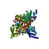
|
|---|---|
| 1 |
|
-Components
-Protein , 1 types, 1 molecules A
| #1: Protein | Mass: 220903.500 Da / Num. of mol.: 1 Source method: isolated from a genetically manipulated source Source: (gene. exp.) Homo sapiens (human) / Gene: SCN10A / Production host: Homo sapiens (human) / References: UniProt: Q9Y5Y9 |
|---|
-Sugars , 2 types, 5 molecules 
| #2: Polysaccharide | 2-acetamido-2-deoxy-beta-D-glucopyranose-(1-4)-2-acetamido-2-deoxy-beta-D-glucopyranose Source method: isolated from a genetically manipulated source |
|---|---|
| #3: Sugar | ChemComp-NAG /  Type: D-saccharide, beta linking / Mass: 221.208 Da / Num. of mol.: 4 / Source method: obtained synthetically / Formula: C8H15NO6 Type: D-saccharide, beta linking / Mass: 221.208 Da / Num. of mol.: 4 / Source method: obtained synthetically / Formula: C8H15NO6 |
-Non-polymers , 4 types, 17 molecules 


| #4: Chemical | ChemComp-CLR /  Mass: 386.654 Da / Num. of mol.: 4 / Source method: obtained synthetically / Formula: C27H46O Mass: 386.654 Da / Num. of mol.: 4 / Source method: obtained synthetically / Formula: C27H46O#5: Chemical |  Mass: 787.121 Da / Num. of mol.: 3 / Source method: obtained synthetically / Formula: C44H85NO8P / Comment: DOPC, phospholipid*YM Mass: 787.121 Da / Num. of mol.: 3 / Source method: obtained synthetically / Formula: C44H85NO8P / Comment: DOPC, phospholipid*YM#6: Chemical | ChemComp-LPE /  Mass: 510.708 Da / Num. of mol.: 8 / Source method: obtained synthetically / Formula: C26H57NO6P Mass: 510.708 Da / Num. of mol.: 8 / Source method: obtained synthetically / Formula: C26H57NO6P#7: Chemical |  Mass: 792.075 Da / Num. of mol.: 2 / Source method: obtained synthetically / Formula: C42H82NO10P Mass: 792.075 Da / Num. of mol.: 2 / Source method: obtained synthetically / Formula: C42H82NO10P |
|---|
-Details
| Has ligand of interest | N |
|---|---|
| Has protein modification | Y |
-Experimental details
-Experiment
| Experiment | Method: ELECTRON MICROSCOPY |
|---|---|
| EM experiment | Aggregation state: PARTICLE / 3D reconstruction method: single particle reconstruction |
- Sample preparation
| Component | Name: sodium channel II / Type: COMPLEX / Entity ID: #1 / Source: RECOMBINANT |
|---|---|
| Source (natural) | Organism: Homo sapiens (human) |
| Source (recombinant) | Organism: Homo sapiens (human) |
| Buffer solution | pH: 7.5 |
| Specimen | Embedding applied: NO / Shadowing applied: NO / Staining applied: NO / Vitrification applied: YES |
| Vitrification | Cryogen name: ETHANE |
- Electron microscopy imaging
| Experimental equipment |  Model: Titan Krios / Image courtesy: FEI Company |
|---|---|
| Microscopy | Model: FEI TITAN KRIOS |
| Electron gun | Electron source:  FIELD EMISSION GUN / Accelerating voltage: 300 kV / Illumination mode: FLOOD BEAM FIELD EMISSION GUN / Accelerating voltage: 300 kV / Illumination mode: FLOOD BEAM |
| Electron lens | Mode: BRIGHT FIELD / Nominal defocus max: 1800 nm / Nominal defocus min: 1500 nm |
| Image recording | Electron dose: 50 e/Å2 / Film or detector model: GATAN K3 (6k x 4k) |
- Processing
| Software | Name: PHENIX / Version: 1.18_3855: / Classification: refinement | ||||||||||||||||||||||||
|---|---|---|---|---|---|---|---|---|---|---|---|---|---|---|---|---|---|---|---|---|---|---|---|---|---|
| EM software | Name: PHENIX / Category: model refinement | ||||||||||||||||||||||||
| CTF correction | Type: PHASE FLIPPING AND AMPLITUDE CORRECTION | ||||||||||||||||||||||||
| 3D reconstruction | Resolution: 3.1 Å / Resolution method: FSC 0.143 CUT-OFF / Num. of particles: 410478 / Symmetry type: POINT | ||||||||||||||||||||||||
| Refine LS restraints |
|
