 Movie
Movie Controller


+ Open data

- Basic information
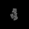
| Entry | Database: PDB / ID: 7uvl | |||||||||||||||||||||||||||||||||||||||
|---|---|---|---|---|---|---|---|---|---|---|---|---|---|---|---|---|---|---|---|---|---|---|---|---|---|---|---|---|---|---|---|---|---|---|---|---|---|---|---|---|
| Title | IgA1 Protease with IgA1 substrate | |||||||||||||||||||||||||||||||||||||||
 Components Components |
| |||||||||||||||||||||||||||||||||||||||
 Keywords Keywords | IMMUNE SYSTEM / complex | |||||||||||||||||||||||||||||||||||||||
| Function / homology |  Function and homology information Function and homology informationsecretory dimeric IgA immunoglobulin complex / monomeric IgA immunoglobulin complex / secretory IgA immunoglobulin complex / IgA immunoglobulin complex / glomerular filtration / immunoglobulin complex, circulating / IgG immunoglobulin complex / positive regulation of respiratory burst / complement activation, classical pathway / Scavenging of heme from plasma ...secretory dimeric IgA immunoglobulin complex / monomeric IgA immunoglobulin complex / secretory IgA immunoglobulin complex / IgA immunoglobulin complex / glomerular filtration / immunoglobulin complex, circulating / IgG immunoglobulin complex / positive regulation of respiratory burst / complement activation, classical pathway / Scavenging of heme from plasma / antigen binding / serine-type peptidase activity / Cell surface interactions at the vascular wall / B cell receptor signaling pathway / metalloendopeptidase activity / antibacterial humoral response / blood microparticle / adaptive immune response / immune response / : / extracellular exosome / extracellular region / zinc ion binding / membrane / plasma membrane Similarity search - Function | |||||||||||||||||||||||||||||||||||||||
| Biological species |  Gemella haemolysans (bacteria) Gemella haemolysans (bacteria) Homo sapiens (human) Homo sapiens (human) | |||||||||||||||||||||||||||||||||||||||
| Method | ELECTRON MICROSCOPY / single particle reconstruction / cryo EM / Resolution: 3.56 Å | |||||||||||||||||||||||||||||||||||||||
 Authors Authors | Eisenmesser, Z.E. / Zheng, H. | |||||||||||||||||||||||||||||||||||||||
| Funding support |  United States, 1items United States, 1items
| |||||||||||||||||||||||||||||||||||||||
 Citation Citation | Journal: Commun Biol / Year: 2022 Title: A substrate-induced gating mechanism is conserved among Gram-positive IgA1 metalloproteases. Authors: Jasmina S Redzic / Jeremy Rahkola / Norman Tran / Todd Holyoak / Eunjeong Lee / Antonio Javier Martín-Galiano / Nancy Meyer / Hongjin Zheng / Elan Eisenmesser /   Abstract: The mucosal adaptive immune response is dependent on the production of IgA antibodies and particularly IgA1, yet opportunistic bacteria have evolved mechanisms to specifically block this response by ...The mucosal adaptive immune response is dependent on the production of IgA antibodies and particularly IgA1, yet opportunistic bacteria have evolved mechanisms to specifically block this response by producing IgA1 proteases (IgA1Ps). Our lab was the first to describe the structures of a metal-dependent IgA1P (metallo-IgA1P) produced from Gram-positive Streptococcus pneumoniae both in the absence and presence of its IgA1 substrate through cryo-EM single particle reconstructions. This prior study revealed an active-site gating mechanism reliant on substrate-induced conformational changes to the enzyme that begged the question of whether such a mechanism is conserved among the wider Gram-positive metallo-IgA1P subfamily of virulence factors. Here, we used cryo-EM to characterize the metallo-IgA1P of a more distantly related family member from Gemella haemolysans, an emerging opportunistic pathogen implicated in meningitis, endocarditis, and more recently bacteremia in the elderly. While the substrate-free structures of these two metallo-IgA1Ps exhibit differences in the relative starting positions of the domain responsible for gating substrate, the enzymes have similar domain orientations when bound to IgA1. Together with biochemical studies that indicate these metallo-IgA1Ps have similar binding affinities and activities, these data indicate that metallo-IgA1P binding requires the specific IgA1 substrate to open the enzymes for access to their active site and thus, largely conform to an "induced fit" model. | |||||||||||||||||||||||||||||||||||||||
| History |
|
- Structure visualization
| Structure viewer | Molecule: MolmilJmol/JSmol |
|---|
- Downloads & links
-Download
| PDBx/mmCIF format | 7uvl.cif.gz | 380.8 KB | Display | PDBx/mmCIF format |
|---|---|---|---|---|
| PDB format | pdb7uvl.ent.gz | Display | PDB format | |
| PDBx/mmJSON format | 7uvl.json.gz | Tree view | PDBx/mmJSON format | |
| Others |  Other downloads Other downloads |
-Validation report
| Arichive directory | https://data.pdbj.org/pub/pdb/validation_reports/uv/7uvlftp://data.pdbj.org/pub/pdb/validation_reports/uv/7uvl | HTTPS FTP |
|---|
-Related structure data
| Related structure data | 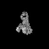 26813MC  7uvkC M: map data used to model this data C: citing same article ( |
|---|---|
| Similar structure data |
-Links
 PDBj
PDBj


- Assembly
| Deposited unit | 
|
|---|---|
| 1 |
|
 gel filtration
-Components
| #1: Protein | Mass: 146820.328 Da / Num. of mol.: 1 / Mutation: A942E Source method: isolated from a genetically manipulated source Source: (gene. exp.) Gemella haemolysans (bacteria) / Gene: GEMHA0001_0491 / Production host: | ||||||
|---|---|---|---|---|---|---|---|
| #2: Protein | Mass: 22784.846 Da / Num. of mol.: 2 Source method: isolated from a genetically manipulated source Source: (gene. exp.) Homo sapiens (human) / Gene: IGHA1 / Production host:  #3: Antibody | | Mass: 24009.725 Da / Num. of mol.: 1 Source method: isolated from a genetically manipulated source Source: (gene. exp.) Homo sapiens (human) / Production host: #4: Antibody | | Mass: 24554.428 Da / Num. of mol.: 1 Source method: isolated from a genetically manipulated source Source: (gene. exp.) Homo sapiens (human) / Production host: Has protein modification | Y | |
-Experimental details
-Experiment
| Experiment | Method: ELECTRON MICROSCOPY |
|---|---|
| EM experiment | Aggregation state: PARTICLE / 3D reconstruction method: single particle reconstruction |
- Sample preparation
| Component | Name: IgA1 Protease with IgA1 substrate / Type: COMPLEX / Entity ID: all / Source: MULTIPLE SOURCES | |||||||||||||||
|---|---|---|---|---|---|---|---|---|---|---|---|---|---|---|---|---|
| Source (natural) |
| |||||||||||||||
| Source (recombinant) |
| |||||||||||||||
| Buffer solution | pH: 7.4 | |||||||||||||||
| Buffer component |
| |||||||||||||||
| Specimen | Embedding applied: NO / Shadowing applied: NO / Staining applied: NO / Vitrification applied: YES | |||||||||||||||
| Vitrification | Cryogen name: ETHANE |
- Electron microscopy imaging
| Experimental equipment |  Model: Titan Krios / Image courtesy: FEI Company |
|---|---|
| Microscopy | Model: FEI TITAN KRIOS |
| Electron gun | Electron source: OTHER / Accelerating voltage: 300 kV / Illumination mode: OTHER |
| Electron lens | Mode: OTHER / Nominal defocus max: 5000 nm / Nominal defocus min: 1200 nm |
| Image recording | Electron dose: 49 e/Å2 / Film or detector model: GATAN K3 (6k x 4k) |
- Processing
| Software | Name: PHENIX / Version: 1.19.2_4158: / Classification: refinement | ||||||||||||||||||||||||
|---|---|---|---|---|---|---|---|---|---|---|---|---|---|---|---|---|---|---|---|---|---|---|---|---|---|
| EM software | Name: PHENIX / Category: model refinement | ||||||||||||||||||||||||
| CTF correction | Type: NONE | ||||||||||||||||||||||||
| 3D reconstruction | Resolution: 3.56 Å / Resolution method: FSC 0.143 CUT-OFF / Num. of particles: 205808 / Symmetry type: POINT | ||||||||||||||||||||||||
| Refine LS restraints |
|
