National Institutes of Health/National Institute Of Allergy and Infectious Diseases (NIH/NIAID)
R21 AI146295
United States
Citation
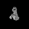
Journal: Commun Biol / Year: 2022 Title: A substrate-induced gating mechanism is conserved among Gram-positive IgA1 metalloproteases. Authors: Jasmina S Redzic / Jeremy Rahkola / Norman Tran / Todd Holyoak / Eunjeong Lee / Antonio Javier Martín-Galiano / Nancy Meyer / Hongjin Zheng / Elan Eisenmesser / Abstract: The mucosal adaptive immune response is dependent on the production of IgA antibodies and particularly IgA1, yet opportunistic bacteria have evolved mechanisms to specifically block this response by ...The mucosal adaptive immune response is dependent on the production of IgA antibodies and particularly IgA1, yet opportunistic bacteria have evolved mechanisms to specifically block this response by producing IgA1 proteases (IgA1Ps). Our lab was the first to describe the structures of a metal-dependent IgA1P (metallo-IgA1P) produced from Gram-positive Streptococcus pneumoniae both in the absence and presence of its IgA1 substrate through cryo-EM single particle reconstructions. This prior study revealed an active-site gating mechanism reliant on substrate-induced conformational changes to the enzyme that begged the question of whether such a mechanism is conserved among the wider Gram-positive metallo-IgA1P subfamily of virulence factors. Here, we used cryo-EM to characterize the metallo-IgA1P of a more distantly related family member from Gemella haemolysans, an emerging opportunistic pathogen implicated in meningitis, endocarditis, and more recently bacteremia in the elderly. While the substrate-free structures of these two metallo-IgA1Ps exhibit differences in the relative starting positions of the domain responsible for gating substrate, the enzymes have similar domain orientations when bound to IgA1. Together with biochemical studies that indicate these metallo-IgA1Ps have similar binding affinities and activities, these data indicate that metallo-IgA1P binding requires the specific IgA1 substrate to open the enzymes for access to their active site and thus, largely conform to an "induced fit" model.
History
Deposition
May 2, 2022
Deposition site: RCSB / Processing site: RCSB
Revision 1.0
Nov 23, 2022
Provider: repository / Type: Initial release
Revision 1.1
Jun 12, 2024
Group: Data collection / Category: chem_comp_atom / chem_comp_bond
In the structure databanks used in Yorodumi, some data are registered as the other names, "COVID-19 virus" and "2019-nCoV". Here are the details of the virus and the list of structure data.
Jan 31, 2019. EMDB accession codes are about to change! (news from PDBe EMDB page)
EMDB accession codes are about to change! (news from PDBe EMDB page)
The allocation of 4 digits for EMDB accession codes will soon come to an end. Whilst these codes will remain in use, new EMDB accession codes will include an additional digit and will expand incrementally as the available range of codes is exhausted. The current 4-digit format prefixed with “EMD-” (i.e. EMD-XXXX) will advance to a 5-digit format (i.e. EMD-XXXXX), and so on. It is currently estimated that the 4-digit codes will be depleted around Spring 2019, at which point the 5-digit format will come into force.
The EM Navigator/Yorodumi systems omit the EMD- prefix.
Related info.:Q: What is EMD? / ID/Accession-code notation in Yorodumi/EM Navigator
Yorodumi is a browser for structure data from EMDB, PDB, SASBDB, etc.
This page is also the successor to EM Navigator detail page, and also detail information page/front-end page for Omokage search.
The word "yorodu" (or yorozu) is an old Japanese word meaning "ten thousand". "mi" (miru) is to see.
Related info.:EMDB / PDB / SASBDB / Comparison of 3 databanks / Yorodumi Search / Aug 31, 2016. New EM Navigator & Yorodumi / Yorodumi Papers / Jmol/JSmol / Function and homology information / Changes in new EM Navigator and Yorodumi
 Movie
Movie Controller


 Open data
 Basic information
 Components
 Keywords
 Function and homology information
 Gemella haemolysans (bacteria)
 Authors
 United States, 1items
  Citation

 Structure visualization
 Downloads & links
 Other downloads
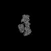

 PDBj
PDBj Assembly
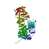
 gel filtration
 Sample preparation
 Electron microscopy imaging
 Processing