 Movie
Movie Controller


+ Open data

- Basic information
| Entry | Database: PDB / ID: 7uph | ||||||||||||||||||||||||||||||||||||||||||||||||||||||
|---|---|---|---|---|---|---|---|---|---|---|---|---|---|---|---|---|---|---|---|---|---|---|---|---|---|---|---|---|---|---|---|---|---|---|---|---|---|---|---|---|---|---|---|---|---|---|---|---|---|---|---|---|---|---|---|
| Title | Structure of a ribosome with tethered subunits | ||||||||||||||||||||||||||||||||||||||||||||||||||||||
 Components Components |
| ||||||||||||||||||||||||||||||||||||||||||||||||||||||
 Keywords Keywords | RIBOSOME / Engineered / Tethered / Synthetic | ||||||||||||||||||||||||||||||||||||||||||||||||||||||
| Function / homology |  Function and homology information Function and homology informationDnaA-L2 complex / negative regulation of DNA-templated DNA replication initiation / cytosolic ribosome assembly / ribosome assembly / assembly of large subunit precursor of preribosome / mRNA 5'-UTR binding / large ribosomal subunit / transferase activity / ribosomal small subunit assembly / ribosome binding ...DnaA-L2 complex / negative regulation of DNA-templated DNA replication initiation / cytosolic ribosome assembly / ribosome assembly / assembly of large subunit precursor of preribosome / mRNA 5'-UTR binding / large ribosomal subunit / transferase activity / ribosomal small subunit assembly / ribosome binding / 5S rRNA binding / ribosomal large subunit assembly / small ribosomal subunit / small ribosomal subunit rRNA binding / cytosolic small ribosomal subunit / large ribosomal subunit rRNA binding / cytosolic large ribosomal subunit / cytoplasmic translation / tRNA binding / negative regulation of translation / rRNA binding / structural constituent of ribosome / ribosome / translation / ribonucleoprotein complex / response to antibiotic / mRNA binding / RNA binding / zinc ion binding / membrane / cytoplasm / cytosol Similarity search - Function | ||||||||||||||||||||||||||||||||||||||||||||||||||||||
| Biological species |  | ||||||||||||||||||||||||||||||||||||||||||||||||||||||
| Method | ELECTRON MICROSCOPY / single particle reconstruction / cryo EM / Resolution: 4.18 Å | ||||||||||||||||||||||||||||||||||||||||||||||||||||||
 Authors Authors | Kim, D.S. / Watkins, A. / Bidstrup, E. / Lee, J. / Topkar, V.V. / Kofman, C. / Schwarz, K.J. / Liu, Y. / Pintilie, G. / Roney, E. ...Kim, D.S. / Watkins, A. / Bidstrup, E. / Lee, J. / Topkar, V.V. / Kofman, C. / Schwarz, K.J. / Liu, Y. / Pintilie, G. / Roney, E. / Das, R. / Jewett, M.C. | ||||||||||||||||||||||||||||||||||||||||||||||||||||||
| Funding support |  United States, 4items United States, 4items
| ||||||||||||||||||||||||||||||||||||||||||||||||||||||
 Citation Citation | Journal: Nat Chem Biol / Year: 2022 Title: Three-dimensional structure-guided evolution of a ribosome with tethered subunits. Authors: Do Soon Kim / Andrew Watkins / Erik Bidstrup / Joongoo Lee / Ved Topkar / Camila Kofman / Kevin J Schwarz / Yan Liu / Grigore Pintilie / Emily Roney / Rhiju Das / Michael C Jewett /  Abstract: RNA-based macromolecular machines, such as the ribosome, have functional parts reliant on structural interactions spanning sequence-distant regions. These features limit evolutionary exploration of ...RNA-based macromolecular machines, such as the ribosome, have functional parts reliant on structural interactions spanning sequence-distant regions. These features limit evolutionary exploration of mutant libraries and confound three-dimensional structure-guided design. To address these challenges, we describe Evolink (evolution and linkage), a method that enables high-throughput evolution of sequence-distant regions in large macromolecular machines, and library design guided by computational RNA modeling to enable exploration of structurally stable designs. Using Evolink, we evolved a tethered ribosome with a 58% increased activity in orthogonal protein translation and a 97% improvement in doubling times in SQ171 cells compared to a previously developed tethered ribosome, and reveal new permissible sequences in a pair of ribosomal helices with previously explored biological function. The Evolink approach may enable enhanced engineering of macromolecular machines for new and improved functions for synthetic biology. #1: Journal: Protein Sci / Year: 2021Title: UCSF ChimeraX: Structure visualization for researchers, educators, and developers. Authors: Pettersen, E.F. / Goddard, T.D. / Huang, C.C. / Meng, E.C. / Couch, G.S. / Croll, T.I. / Morris, J.H. / Ferrin, T.E. #2: Journal: Nat Methods / Year: 2017Title: cryoSPARC: algorithms for rapid unsupervised cryo-EM structure determination. Authors: Punjani, A. / Rubinstein, J.L. / Fleet, D.J. / Brubaker, M.A. #3: Journal: Acta Crystallogr D Struct Biol / Year: 2019 Title: Macromolecular structure determination using X-rays, neutrons and electrons: recent developments in Phenix. Authors: Dorothee Liebschner / Pavel V Afonine / Matthew L Baker / Gábor Bunkóczi / Vincent B Chen / Tristan I Croll / Bradley Hintze / Li Wei Hung / Swati Jain / Airlie J McCoy / Nigel W Moriarty ...Authors: Dorothee Liebschner / Pavel V Afonine / Matthew L Baker / Gábor Bunkóczi / Vincent B Chen / Tristan I Croll / Bradley Hintze / Li Wei Hung / Swati Jain / Airlie J McCoy / Nigel W Moriarty / Robert D Oeffner / Billy K Poon / Michael G Prisant / Randy J Read / Jane S Richardson / David C Richardson / Massimo D Sammito / Oleg V Sobolev / Duncan H Stockwell / Thomas C Terwilliger / Alexandre G Urzhumtsev / Lizbeth L Videau / Christopher J Williams / Paul D Adams /   Abstract: Diffraction (X-ray, neutron and electron) and electron cryo-microscopy are powerful methods to determine three-dimensional macromolecular structures, which are required to understand biological ...Diffraction (X-ray, neutron and electron) and electron cryo-microscopy are powerful methods to determine three-dimensional macromolecular structures, which are required to understand biological processes and to develop new therapeutics against diseases. The overall structure-solution workflow is similar for these techniques, but nuances exist because the properties of the reduced experimental data are different. Software tools for structure determination should therefore be tailored for each method. Phenix is a comprehensive software package for macromolecular structure determination that handles data from any of these techniques. Tasks performed with Phenix include data-quality assessment, map improvement, model building, the validation/rebuilding/refinement cycle and deposition. Each tool caters to the type of experimental data. The design of Phenix emphasizes the automation of procedures, where possible, to minimize repetitive and time-consuming manual tasks, while default parameters are chosen to encourage best practice. A graphical user interface provides access to many command-line features of Phenix and streamlines the transition between programs, project tracking and re-running of previous tasks. | ||||||||||||||||||||||||||||||||||||||||||||||||||||||
| History |
|
- Structure visualization
| Structure viewer | Molecule: MolmilJmol/JSmol |
|---|
- Downloads & links
-Download
| PDBx/mmCIF format | 7uph.cif.gz | 3 MB | Display | PDBx/mmCIF format |
|---|---|---|---|---|
| PDB format | pdb7uph.ent.gz | Display | PDB format | |
| PDBx/mmJSON format | 7uph.json.gz | Tree view | PDBx/mmJSON format | |
| Others |  Other downloads Other downloads |
-Validation report
| Arichive directory | https://data.pdbj.org/pub/pdb/validation_reports/up/7uphftp://data.pdbj.org/pub/pdb/validation_reports/up/7uph | HTTPS FTP |
|---|
-Related structure data
| Related structure data |  26666MC M: map data used to model this data C: citing same article ( |
|---|---|
| Similar structure data |
-Links
 PDBj
PDBj


- Assembly
| Deposited unit | 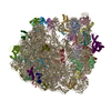
|
|---|---|
| 1 |
|
-Components
-30S ribosomal protein ... , 20 types, 20 molecules ABCDEFGHWXYZabcdefgh
| #1: Protein | Mass: 11475.364 Da / Num. of mol.: 1 Source method: isolated from a genetically manipulated source Source: (gene. exp.) |
|---|---|
| #2: Protein | Mass: 10159.621 Da / Num. of mol.: 1 Source method: isolated from a genetically manipulated source Source: (gene. exp.) |
| #3: Protein | Mass: 9207.572 Da / Num. of mol.: 1 Source method: isolated from a genetically manipulated source Source: (gene. exp.) |
| #4: Protein | Mass: 9263.946 Da / Num. of mol.: 1 Source method: isolated from a genetically manipulated source Source: (gene. exp.) |
| #5: Protein | Mass: 6466.477 Da / Num. of mol.: 1 Source method: isolated from a genetically manipulated source Source: (gene. exp.) |
| #6: Protein | Mass: 9057.626 Da / Num. of mol.: 1 Source method: isolated from a genetically manipulated source Source: (gene. exp.) |
| #7: Protein | Mass: 9506.190 Da / Num. of mol.: 1 Source method: isolated from a genetically manipulated source Source: (gene. exp.) |
| #8: Protein | Mass: 6629.744 Da / Num. of mol.: 1 Source method: isolated from a genetically manipulated source Source: (gene. exp.) |
| #21: Protein | Mass: 24971.764 Da / Num. of mol.: 1 Source method: isolated from a genetically manipulated source Source: (gene. exp.) |
| #22: Protein | Mass: 23078.785 Da / Num. of mol.: 1 Source method: isolated from a genetically manipulated source Source: (gene. exp.) |
| #23: Protein | Mass: 23383.002 Da / Num. of mol.: 1 Source method: isolated from a genetically manipulated source Source: (gene. exp.) |
| #24: Protein | Mass: 15804.282 Da / Num. of mol.: 1 Source method: isolated from a genetically manipulated source Source: (gene. exp.) |
| #25: Protein | Mass: 11669.371 Da / Num. of mol.: 1 Source method: isolated from a genetically manipulated source Source: (gene. exp.) |
| #26: Protein | Mass: 16861.523 Da / Num. of mol.: 1 Source method: isolated from a genetically manipulated source Source: (gene. exp.) |
| #27: Protein | Mass: 14015.361 Da / Num. of mol.: 1 Source method: isolated from a genetically manipulated source Source: (gene. exp.) |
| #28: Protein | Mass: 14554.882 Da / Num. of mol.: 1 Source method: isolated from a genetically manipulated source Source: (gene. exp.) |
| #29: Protein | Mass: 11196.988 Da / Num. of mol.: 1 Source method: isolated from a genetically manipulated source Source: (gene. exp.) |
| #30: Protein | Mass: 12487.200 Da / Num. of mol.: 1 Source method: isolated from a genetically manipulated source Source: (gene. exp.) |
| #31: Protein | Mass: 13683.053 Da / Num. of mol.: 1 Source method: isolated from a genetically manipulated source Source: (gene. exp.) |
| #32: Protein | Mass: 12625.753 Da / Num. of mol.: 1 Source method: isolated from a genetically manipulated source Source: (gene. exp.) |
-RNA chain , 2 types, 2 molecules IJ
| #9: RNA chain | Mass: 1439279.875 Da / Num. of mol.: 1 Source method: isolated from a genetically manipulated source Source: (gene. exp.) |
|---|---|
| #10: RNA chain | Mass: 38177.762 Da / Num. of mol.: 1 Source method: isolated from a genetically manipulated source Source: (gene. exp.) |
+50S ribosomal protein ... , 27 types, 27 molecules KLMNORSTUVijklmnopqrstuvwxy
-Details
| Has ligand of interest | N |
|---|---|
| Has protein modification | Y |
-Experimental details
-Experiment
| Experiment | Method: ELECTRON MICROSCOPY |
|---|---|
| EM experiment | Aggregation state: PARTICLE / 3D reconstruction method: single particle reconstruction |
- Sample preparation
| Component | Name: Ribosome with tethered subunits / Type: RIBOSOME / Entity ID: all / Source: RECOMBINANT |
|---|---|
| Molecular weight | Experimental value: NO |
| Source (natural) | Organism: |
| Source (recombinant) | Organism: |
| Buffer solution | pH: 7.5 |
| Specimen | Embedding applied: NO / Shadowing applied: NO / Staining applied: NO / Vitrification applied: YES |
| Specimen support | Grid material: COPPER / Grid type: Quantifoil R2/1 |
| Vitrification | Cryogen name: ETHANE |
- Electron microscopy imaging
| Microscopy | Model: TFS TALOS |
|---|---|
| Electron gun | Electron source:  FIELD EMISSION GUN / Accelerating voltage: 200 kV / Illumination mode: SPOT SCAN FIELD EMISSION GUN / Accelerating voltage: 200 kV / Illumination mode: SPOT SCAN |
| Electron lens | Mode: BRIGHT FIELD / Nominal defocus max: 2200 nm / Nominal defocus min: 1200 nm |
| Image recording | Average exposure time: 1.35 sec. / Electron dose: 50 e/Å2 / Film or detector model: GATAN K3 (6k x 4k) |
- Processing
| Software | Name: UCSF ChimeraX / Version: 1.3/v9 / Classification: model building / URL: https://www.rbvi.ucsf.edu/chimerax/ / Os: macOS / Type: package | ||||||||||||||||||||||||||||
|---|---|---|---|---|---|---|---|---|---|---|---|---|---|---|---|---|---|---|---|---|---|---|---|---|---|---|---|---|---|
| EM software |
| ||||||||||||||||||||||||||||
| CTF correction | Type: NONE | ||||||||||||||||||||||||||||
| Particle selection | Num. of particles selected: 102279 | ||||||||||||||||||||||||||||
| 3D reconstruction | Resolution: 4.18 Å / Resolution method: FSC 0.143 CUT-OFF / Num. of particles: 34852 / Symmetry type: POINT | ||||||||||||||||||||||||||||
| Atomic model building | Protocol: AB INITIO MODEL | ||||||||||||||||||||||||||||
| Atomic model building | PDB-ID: 4YBB Accession code: 4YBB / Source name: PDB / Type: experimental model |

