Movie
Movie Controller


+ Open data

- Basic information
| Entry |  | |||||||||||||||
|---|---|---|---|---|---|---|---|---|---|---|---|---|---|---|---|---|
| Title | Structure of a ribosome with tethered subunits | |||||||||||||||
 Map data Map data | Cryo-EM structure of the RiboTv3 tethered ribosome. Raw micrograph acquisition was done with a Talos Arctica (200kV) microscope at 100,000X magnification. Map generation was done with CryoSPARC. | |||||||||||||||
 Sample Sample |
| |||||||||||||||
 Keywords Keywords | Engineered / Tethered / Synthetic / RIBOSOME | |||||||||||||||
| Function / homology |  Function and homology information Function and homology informationDnaA-L2 complex / negative regulation of DNA-templated DNA replication initiation / cytosolic ribosome assembly / ribosome assembly / assembly of large subunit precursor of preribosome / mRNA 5'-UTR binding / large ribosomal subunit / transferase activity / ribosomal small subunit assembly / ribosome binding ...DnaA-L2 complex / negative regulation of DNA-templated DNA replication initiation / cytosolic ribosome assembly / ribosome assembly / assembly of large subunit precursor of preribosome / mRNA 5'-UTR binding / large ribosomal subunit / transferase activity / ribosomal small subunit assembly / ribosome binding / 5S rRNA binding / ribosomal large subunit assembly / small ribosomal subunit / small ribosomal subunit rRNA binding / cytosolic small ribosomal subunit / large ribosomal subunit rRNA binding / cytosolic large ribosomal subunit / cytoplasmic translation / tRNA binding / negative regulation of translation / rRNA binding / structural constituent of ribosome / ribosome / translation / ribonucleoprotein complex / response to antibiotic / mRNA binding / RNA binding / zinc ion binding / membrane / cytoplasm / cytosol Similarity search - Function | |||||||||||||||
| Biological species |  | |||||||||||||||
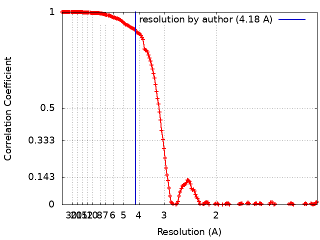
| Method | single particle reconstruction / cryo EM / Resolution: 4.18 Å | |||||||||||||||
 Authors Authors | Kim DS / Watkins A / Bidstrup E / Lee J / Topkar VV / Kofman C / Schwarz KJ / Liu Y / Pintilie G / Roney E ...Kim DS / Watkins A / Bidstrup E / Lee J / Topkar VV / Kofman C / Schwarz KJ / Liu Y / Pintilie G / Roney E / Das R / Jewett MC | |||||||||||||||
| Funding support |  United States, 4 items United States, 4 items
| |||||||||||||||
 Citation Citation | Journal: Acta Crystallogr D Struct Biol / Year: 2019 Title: Macromolecular structure determination using X-rays, neutrons and electrons: recent developments in Phenix. Authors: Dorothee Liebschner / Pavel V Afonine / Matthew L Baker / Gábor Bunkóczi / Vincent B Chen / Tristan I Croll / Bradley Hintze / Li Wei Hung / Swati Jain / Airlie J McCoy / Nigel W Moriarty ...Authors: Dorothee Liebschner / Pavel V Afonine / Matthew L Baker / Gábor Bunkóczi / Vincent B Chen / Tristan I Croll / Bradley Hintze / Li Wei Hung / Swati Jain / Airlie J McCoy / Nigel W Moriarty / Robert D Oeffner / Billy K Poon / Michael G Prisant / Randy J Read / Jane S Richardson / David C Richardson / Massimo D Sammito / Oleg V Sobolev / Duncan H Stockwell / Thomas C Terwilliger / Alexandre G Urzhumtsev / Lizbeth L Videau / Christopher J Williams / Paul D Adams /   Abstract: Diffraction (X-ray, neutron and electron) and electron cryo-microscopy are powerful methods to determine three-dimensional macromolecular structures, which are required to understand biological ...Diffraction (X-ray, neutron and electron) and electron cryo-microscopy are powerful methods to determine three-dimensional macromolecular structures, which are required to understand biological processes and to develop new therapeutics against diseases. The overall structure-solution workflow is similar for these techniques, but nuances exist because the properties of the reduced experimental data are different. Software tools for structure determination should therefore be tailored for each method. Phenix is a comprehensive software package for macromolecular structure determination that handles data from any of these techniques. Tasks performed with Phenix include data-quality assessment, map improvement, model building, the validation/rebuilding/refinement cycle and deposition. Each tool caters to the type of experimental data. The design of Phenix emphasizes the automation of procedures, where possible, to minimize repetitive and time-consuming manual tasks, while default parameters are chosen to encourage best practice. A graphical user interface provides access to many command-line features of Phenix and streamlines the transition between programs, project tracking and re-running of previous tasks. #1: Journal: Protein Sci / Year: 2021Title: UCSF ChimeraX: Structure visualization for researchers, educators, and developers. Authors: Pettersen EF / Goddard TD / Huang CC / Meng EC / Couch GS / Croll TI / Morris JH / Ferrin TE #2: Journal: Nat Methods / Year: 2017Title: cryoSPARC: algorithms for rapid unsupervised cryo-EM structure determination. Authors: Punjani A / Rubinstein JL / Fleet DJ / Brubaker MA | |||||||||||||||
| History |
|
- Structure visualization
| Supplemental images |
|---|

- Downloads & links
-EMDB archive
| Map data | emd_26666.map.gz | 236.1 MB | EMDB map data format | |
|---|---|---|---|---|
| Header (meta data) | emd-26666-v30.xmlemd-26666.xml | 83.5 KB 83.5 KB | Display Display | EMDB header |
| FSC (resolution estimation) | emd_26666_fsc.xml | 17.4 KB | Display | FSC data file |
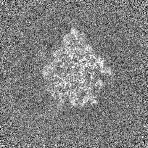
| Images | 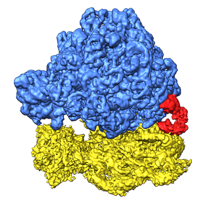 emd_26666.png emd_26666.png | 216.8 KB | ||
| Filedesc metadata | emd-26666.cif.gz | 16.3 KB | ||
| Others | emd_26666_half_map_1.map.gzemd_26666_half_map_2.map.gz | 441.9 MB 441.9 MB | ||
| Archive directory |  http://ftp.pdbj.org/pub/emdb/structures/EMD-26666ftp://ftp.pdbj.org/pub/emdb/structures/EMD-26666 http://ftp.pdbj.org/pub/emdb/structures/EMD-26666ftp://ftp.pdbj.org/pub/emdb/structures/EMD-26666 | HTTPS FTP |
-Related structure data
| Related structure data |  7uphMC M: atomic model generated by this map C: citing same article ( |
|---|---|
| Similar structure data |
-Links
| EMDB pages | EMDB (EBI/PDBe) / EMDataResource |
|---|---|
| Related items in Molecule of the Month |


-Map
| File | Download / File: emd_26666.map.gz / Format: CCP4 / Size: 476.8 MB / Type: IMAGE STORED AS FLOATING POINT NUMBER (4 BYTES) | ||||||||||||||||||||||||||||||||||||
|---|---|---|---|---|---|---|---|---|---|---|---|---|---|---|---|---|---|---|---|---|---|---|---|---|---|---|---|---|---|---|---|---|---|---|---|---|---|
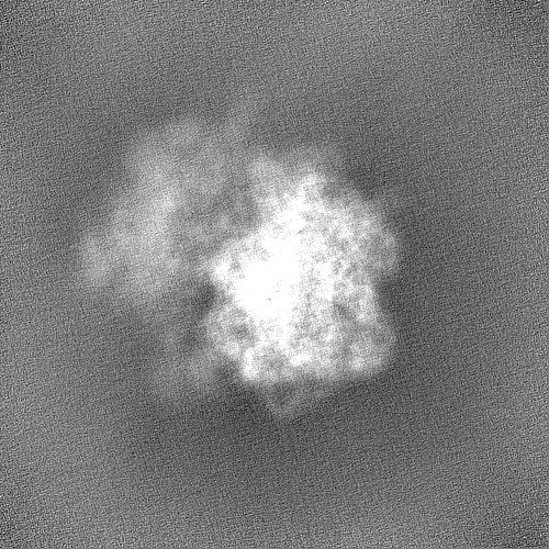
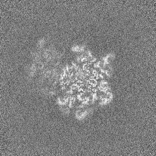
| Annotation | Cryo-EM structure of the RiboTv3 tethered ribosome. Raw micrograph acquisition was done with a Talos Arctica (200kV) microscope at 100,000X magnification. Map generation was done with CryoSPARC. | ||||||||||||||||||||||||||||||||||||
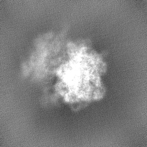
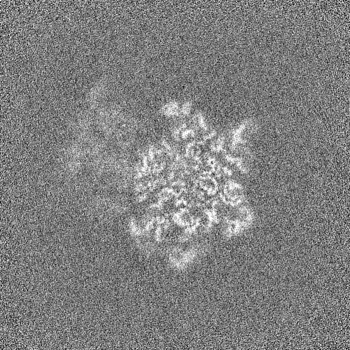
| Projections & slices | Image control
Images are generated by Spider. | ||||||||||||||||||||||||||||||||||||
| Voxel size | X=Y=Z: 0.86 Å | ||||||||||||||||||||||||||||||||||||
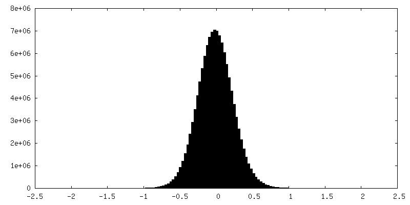
| Density |
| ||||||||||||||||||||||||||||||||||||
| Symmetry | Space group: 1 | ||||||||||||||||||||||||||||||||||||
| Details | EMDB XML:
|
 Z (Sec.)
 Y (Row.)
 X (Col.)


-Supplemental data
-Half map: D 1000263105 em-volume Half Map A
| File | emd_26666_half_map_1.map | ||||||||||||
|---|---|---|---|---|---|---|---|---|---|---|---|---|---|
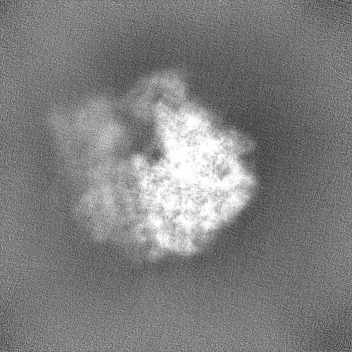
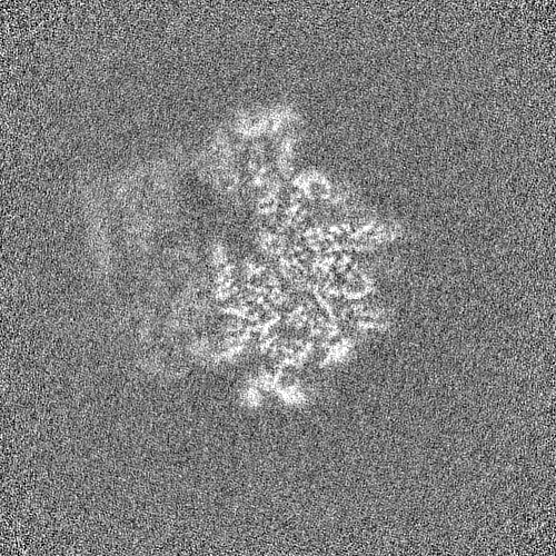
| Annotation | D_1000263105_em-volume Half Map A | ||||||||||||
| Projections & Slices |
| ||||||||||||
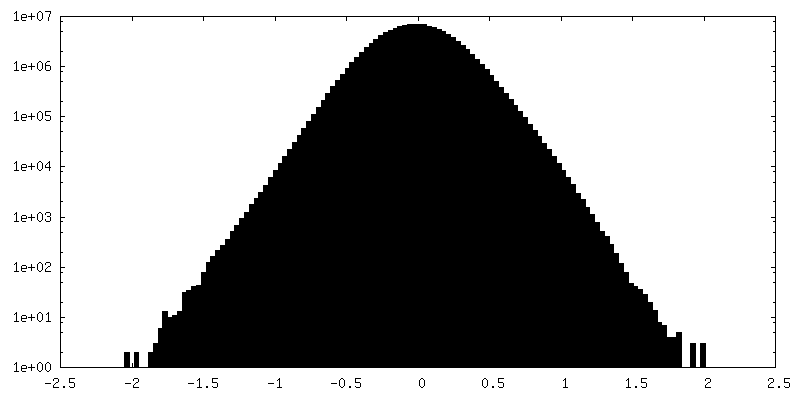
| Density Histograms |


-Half map: D 1000263105 em-volume Half Map B
| File | emd_26666_half_map_2.map | ||||||||||||
|---|---|---|---|---|---|---|---|---|---|---|---|---|---|
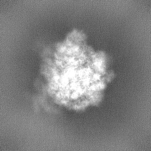
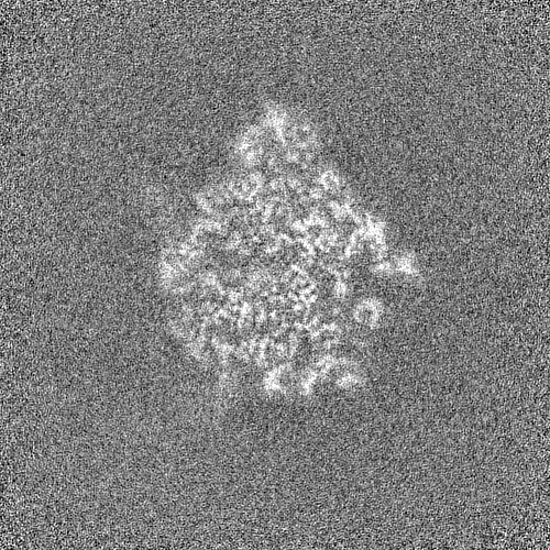
| Annotation | D_1000263105_em-volume Half Map B | ||||||||||||
| Projections & Slices |
| ||||||||||||
| Density Histograms |
- Sample components
+Entire : Ribosome with tethered subunits
+Supramolecule #1: Ribosome with tethered subunits
+Macromolecule #1: 30S ribosomal protein S14
+Macromolecule #2: 30S ribosomal protein S15
+Macromolecule #3: 30S ribosomal protein S16
+Macromolecule #4: 30S ribosomal protein S17
+Macromolecule #5: 30S ribosomal protein S18
+Macromolecule #6: 30S ribosomal protein S19
+Macromolecule #7: 30S ribosomal protein S20
+Macromolecule #8: 30S ribosomal protein S21
+Macromolecule #11: 50S ribosomal protein L2
+Macromolecule #12: 50S ribosomal protein L3
+Macromolecule #13: 50S ribosomal protein L4
+Macromolecule #14: 50S ribosomal protein L5
+Macromolecule #15: 50S ribosomal protein L6
+Macromolecule #16: 50S ribosomal protein L13
+Macromolecule #17: 50S ribosomal protein L14
+Macromolecule #18: 50S ribosomal protein L15
+Macromolecule #19: 50S ribosomal protein L16
+Macromolecule #20: 50S ribosomal protein L17
+Macromolecule #21: 30S ribosomal protein S2
+Macromolecule #22: 30S ribosomal protein S3
+Macromolecule #23: 30S ribosomal protein S4
+Macromolecule #24: 30S ribosomal protein S5
+Macromolecule #25: 30S ribosomal protein S6
+Macromolecule #26: 30S ribosomal protein S7
+Macromolecule #27: 30S ribosomal protein S8
+Macromolecule #28: 30S ribosomal protein S9
+Macromolecule #29: 30S ribosomal protein S10
+Macromolecule #30: 30S ribosomal protein S11
+Macromolecule #31: 30S ribosomal protein S12
+Macromolecule #32: 30S ribosomal protein S13
+Macromolecule #33: 50S ribosomal protein L32
+Macromolecule #34: 50S ribosomal protein L33
+Macromolecule #35: 50S ribosomal protein L34
+Macromolecule #36: 50S ribosomal protein L35
+Macromolecule #37: 50S ribosomal protein L36
+Macromolecule #38: 50S ribosomal protein L18
+Macromolecule #39: 50S ribosomal protein L19
+Macromolecule #40: 50S ribosomal protein L20
+Macromolecule #41: 50S ribosomal protein L21
+Macromolecule #42: 50S ribosomal protein L22
+Macromolecule #43: 50S ribosomal protein L23
+Macromolecule #44: 50S ribosomal protein L24
+Macromolecule #45: 50S ribosomal protein L25
+Macromolecule #46: 50S ribosomal protein L27
+Macromolecule #47: 50S ribosomal protein L28
+Macromolecule #48: 50S ribosomal protein L29
+Macromolecule #49: 50S ribosomal protein L30
+Macromolecule #9: Tethered rRNA
+Macromolecule #10: 5S rRNA
-Experimental details
-Structure determination
| Method | cryo EM |
|---|---|
 Processing Processing | single particle reconstruction |
| Aggregation state | particle |
-Sample preparation
| Buffer | pH: 7.5 |
|---|---|
| Grid | Model: Quantifoil R2/1 / Material: COPPER / Pretreatment - Type: GLOW DISCHARGE |
| Vitrification | Cryogen name: ETHANE |
- Electron microscopy
| Microscope | TFS TALOS |
|---|---|
| Image recording | Film or detector model: GATAN K3 (6k x 4k) / Average exposure time: 1.35 sec. / Average electron dose: 50.0 e/Å2 |
| Electron beam | Acceleration voltage: 200 kV / Electron source:  FIELD EMISSION GUN FIELD EMISSION GUN |
| Electron optics | Illumination mode: SPOT SCAN / Imaging mode: BRIGHT FIELD / Nominal defocus max: 2.2 µm / Nominal defocus min: 1.2 µm |