 Movie
Movie Controller

[English] 日本語
 Yorodumi
- PDB-7tyv: Structure of Lassa Virus glycoprotein (Josiah) bound to Fab 25.10C -
+ Open data

- Basic information
| Entry | Database: PDB / ID: 7tyv | ||||||
|---|---|---|---|---|---|---|---|
| Title | Structure of Lassa Virus glycoprotein (Josiah) bound to Fab 25.10C | ||||||
 Components Components |
| ||||||
 Keywords Keywords | VIRAL PROTEIN/IMMUNE SYSTEM / Surface glycoprotein / antibody Fab fragment / antibody-mediated neutralization / epitope-mapping / VIRAL PROTEIN / VIRAL PROTEIN-IMMUNE SYSTEM complex | ||||||
| Function / homology |  Function and homology information Function and homology informationhost cell Golgi membrane / receptor-mediated endocytosis of virus by host cell / host cell endoplasmic reticulum membrane / fusion of virus membrane with host endosome membrane / viral envelope / virion attachment to host cell / host cell plasma membrane / virion membrane / membrane / metal ion binding Similarity search - Function | ||||||
| Biological species |  Lassa virus Lassa virus Homo sapiens (human) Homo sapiens (human) | ||||||
| Method | ELECTRON MICROSCOPY / single particle reconstruction / cryo EM / Resolution: 2.8 Å | ||||||
 Authors Authors | Enriquez, A.S. / Hastie, K.M. | ||||||
| Funding support |  United States, 1items United States, 1items
| ||||||
 Citation Citation | Journal: Cell Rep / Year: 2022 Title: Delineating the mechanism of anti-Lassa virus GPC-A neutralizing antibodies. Authors: Adrian S Enriquez / Tierra K Buck / Haoyang Li / Michael J Norris / Alex Moon-Walker / Michelle A Zandonatti / Stephanie S Harkins / James E Robinson / Luis M Branco / Robert F Garry / Erica ...Authors: Adrian S Enriquez / Tierra K Buck / Haoyang Li / Michael J Norris / Alex Moon-Walker / Michelle A Zandonatti / Stephanie S Harkins / James E Robinson / Luis M Branco / Robert F Garry / Erica Ollmann Saphire / Kathryn M Hastie / Abstract: Lassa virus (LASV) is the etiologic agent of Lassa Fever, a hemorrhagic disease that is endemic to West Africa. During LASV infection, LASV glycoprotein (GP) engages with multiple host receptors for ...Lassa virus (LASV) is the etiologic agent of Lassa Fever, a hemorrhagic disease that is endemic to West Africa. During LASV infection, LASV glycoprotein (GP) engages with multiple host receptors for cell entry. Neutralizing antibodies against GP are rare and principally target quaternary epitopes displayed only on the metastable, pre-fusion conformation of GP. Currently, the structural features of the neutralizing GPC-A antibody competition group are understudied. Structures of two GPC-A antibodies presented here demonstrate that they bind the side of the pre-fusion GP trimer, bridging the GP1 and GP2 subunits. Complementary biochemical analyses indicate that antibody 25.10C, which is broadly specific, neutralizes by inhibiting binding of the endosomal receptor LAMP1 and also by blocking membrane fusion. The other GPC-A antibody, 36.1F, which is lineage-specific, prevents LAMP1 association only. These data illuminate a site of vulnerability on LASV GP and will guide efforts to elicit broadly reactive therapeutics and vaccines. | ||||||
| History |
|
- Structure visualization
| Structure viewer | Molecule: MolmilJmol/JSmol |
|---|
- Downloads & links
-Download
| PDBx/mmCIF format | 7tyv.cif.gz | 392.5 KB | Display | PDBx/mmCIF format |
|---|---|---|---|---|
| PDB format | pdb7tyv.ent.gz | 320.5 KB | Display | PDB format |
| PDBx/mmJSON format | 7tyv.json.gz | Tree view | PDBx/mmJSON format | |
| Others |  Other downloads Other downloads |
-Validation report
| Arichive directory | https://data.pdbj.org/pub/pdb/validation_reports/ty/7tyvftp://data.pdbj.org/pub/pdb/validation_reports/ty/7tyv | HTTPS FTP |
|---|
-Related structure data
| Related structure data | 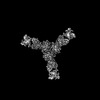 26195MC 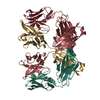 7s8gC  7s8hC M: map data used to model this data C: citing same article ( |
|---|---|
| Similar structure data |
-Links
 PDBj
PDBj

- Assembly
| Deposited unit | 
|
|---|---|
| 1 |
|
-Components
-Glycoprotein ... , 2 types, 6 molecules ABCabc
| #1: Protein | Mass: 29098.430 Da / Num. of mol.: 3 / Mutation: R207C, Residues 256-259 mutated from RRLL to RRRR Source method: isolated from a genetically manipulated source Source: (gene. exp.) Lassa virus / Strain: Mouse/Sierra Leone/Josiah/1976 / Gene: GPC, GP-C / Production host:  #4: Protein | Mass: 19116.732 Da / Num. of mol.: 3 / Mutation: E329P, M332T, G360C Source method: isolated from a genetically manipulated source Source: (gene. exp.) Lassa virus / Strain: Mouse/Sierra Leone/Josiah/1976 / Gene: GPC, GP-C / Production host: |
|---|
-Antibody , 2 types, 6 molecules DGHELK
| #2: Antibody | Mass: 24270.250 Da / Num. of mol.: 3 Source method: isolated from a genetically manipulated source Source: (gene. exp.) Homo sapiens (human) / Production host:   Cricetulus griseus (Chinese hamster) Cricetulus griseus (Chinese hamster)#3: Antibody | Mass: 25501.457 Da / Num. of mol.: 3 Source method: isolated from a genetically manipulated source Source: (gene. exp.) Homo sapiens (human) / Production host: Cricetulus griseus (Chinese hamster) |
|---|
-Sugars , 6 types, 25 molecules 
| #5: Polysaccharide | Source method: isolated from a genetically manipulated source #6: Polysaccharide | 2-acetamido-2-deoxy-beta-D-glucopyranose-(1-4)-2-acetamido-2-deoxy-beta-D-glucopyranose Source method: isolated from a genetically manipulated source #7: Polysaccharide | alpha-D-mannopyranose-(1-2)-alpha-D-mannopyranose-(1-3)-beta-D-mannopyranose-(1-4)-2-acetamido-2- ...alpha-D-mannopyranose-(1-2)-alpha-D-mannopyranose-(1-3)-beta-D-mannopyranose-(1-4)-2-acetamido-2-deoxy-beta-D-glucopyranose-(1-4)-2-acetamido-2-deoxy-beta-D-glucopyranose | Source method: isolated from a genetically manipulated source #8: Polysaccharide | Source method: isolated from a genetically manipulated source #9: Polysaccharide | beta-D-mannopyranose-(1-4)-2-acetamido-2-deoxy-beta-D-glucopyranose-(1-4)-2-acetamido-2-deoxy-beta- ...beta-D-mannopyranose-(1-4)-2-acetamido-2-deoxy-beta-D-glucopyranose-(1-4)-2-acetamido-2-deoxy-beta-D-glucopyranose | Source method: isolated from a genetically manipulated source #10: Sugar | ChemComp-NAG /  Type: D-saccharide, beta linking / Mass: 221.208 Da / Num. of mol.: 13 Type: D-saccharide, beta linking / Mass: 221.208 Da / Num. of mol.: 13Source method: isolated from a genetically manipulated source Formula: C8H15NO6 / Feature type: SUBJECT OF INVESTIGATION |
|---|
-Details
| Has ligand of interest | Y |
|---|---|
| Has protein modification | Y |
-Experimental details
-Experiment
| Experiment | Method: ELECTRON MICROSCOPY |
|---|---|
| EM experiment | Aggregation state: PARTICLE / 3D reconstruction method: single particle reconstruction |
- Sample preparation
| Component |
| ||||||||||||||||||||||||
|---|---|---|---|---|---|---|---|---|---|---|---|---|---|---|---|---|---|---|---|---|---|---|---|---|---|
| Molecular weight | Value: 0.35 MDa / Experimental value: YES | ||||||||||||||||||||||||
| Source (natural) |
| ||||||||||||||||||||||||
| Source (recombinant) |
| ||||||||||||||||||||||||
| Buffer solution | pH: 7.4 / Details: pH 7.4 | ||||||||||||||||||||||||
| Buffer component |
| ||||||||||||||||||||||||
| Specimen | Conc.: 0.7 mg/ml / Embedding applied: NO / Shadowing applied: NO / Staining applied: NO / Vitrification applied: YES / Details: This sample was monodisperse | ||||||||||||||||||||||||
| Vitrification | Instrument: FEI VITROBOT MARK II / Cryogen name: ETHANE / Humidity: 100 % / Chamber temperature: 298 K |
- Electron microscopy imaging
| Experimental equipment |  Model: Titan Krios / Image courtesy: FEI Company |
|---|---|
| Microscopy | Model: FEI TITAN KRIOS |
| Electron gun | Electron source:  FIELD EMISSION GUN / Accelerating voltage: 300 kV / Illumination mode: FLOOD BEAM FIELD EMISSION GUN / Accelerating voltage: 300 kV / Illumination mode: FLOOD BEAM |
| Electron lens | Mode: BRIGHT FIELD / Nominal defocus max: 2500 nm / Nominal defocus min: 1000 nm |
| Image recording | Electron dose: 55 e/Å2 / Detector mode: SUPER-RESOLUTION / Film or detector model: GATAN K2 SUMMIT (4k x 4k) |
- Processing
| Software |
| ||||||||||||||||||||||||
|---|---|---|---|---|---|---|---|---|---|---|---|---|---|---|---|---|---|---|---|---|---|---|---|---|---|
| EM software | Name: cryoSPARC / Version: V2.14.2 / Category: 3D reconstruction | ||||||||||||||||||||||||
| CTF correction | Type: PHASE FLIPPING AND AMPLITUDE CORRECTION | ||||||||||||||||||||||||
| Symmetry | Point symmetry: C3 (3 fold cyclic) | ||||||||||||||||||||||||
| 3D reconstruction | Resolution: 2.8 Å / Resolution method: FSC 0.143 CUT-OFF / Num. of particles: 1150349 / Symmetry type: POINT | ||||||||||||||||||||||||
| Refinement | Cross valid method: NONE Stereochemistry target values: GeoStd + Monomer Library + CDL v1.2 | ||||||||||||||||||||||||
| Displacement parameters | Biso mean: 68.47 Å2 | ||||||||||||||||||||||||
| Refine LS restraints |
|
