 Movie
Movie Controller

[English] 日本語
 Yorodumi
- PDB-7tmc: TMEM106B singlet filament extracted from MSTD neurodegenerative h... -
+ Open data

- Basic information
| Entry | Database: PDB / ID: 7tmc | |||||||||
|---|---|---|---|---|---|---|---|---|---|---|
| Title | TMEM106B singlet filament extracted from MSTD neurodegenerative human brain | |||||||||
 Components Components | Transmembrane protein 106B | |||||||||
 Keywords Keywords | NEUROPEPTIDE / TMEM106B / TMEM filament / MSTD | |||||||||
| Function / homology |  Function and homology information Function and homology informationlysosomal protein catabolic process / lysosomal lumen acidification / lysosome localization / regulation of lysosome organization / positive regulation of dendrite development / lysosomal transport / dendrite morphogenesis / lysosome organization / neuron cellular homeostasis / late endosome membrane ...lysosomal protein catabolic process / lysosomal lumen acidification / lysosome localization / regulation of lysosome organization / positive regulation of dendrite development / lysosomal transport / dendrite morphogenesis / lysosome organization / neuron cellular homeostasis / late endosome membrane / ATPase binding / lysosome / endosome / lysosomal membrane / plasma membrane Similarity search - Function | |||||||||
| Biological species |  Homo sapiens (human) Homo sapiens (human) | |||||||||
| Method | ELECTRON MICROSCOPY / helical reconstruction / cryo EM / Resolution: 3.25 Å | |||||||||
 Authors Authors | Hoq, M.R. / Bharath, S.R. / Jiang, W. | |||||||||
| Funding support |  United States, 1items United States, 1items
| |||||||||
 Citation Citation | Journal: Acta Neuropathol / Year: 2023 Title: Cross-β helical filaments of Tau and TMEM106B in gray and white matter of multiple system tauopathy with presenile dementia. Authors: Md Rejaul Hoq / Sakshibeedu R Bharath / Grace I Hallinan / Anllely Fernandez / Frank S Vago / Kadir A Ozcan / Daoyi Li / Holly J Garringer / Ruben Vidal / Bernardino Ghetti / Wen Jiang / | |||||||||
| History |
|
- Structure visualization
| Structure viewer | Molecule: MolmilJmol/JSmol |
|---|
- Downloads & links
-Download
| PDBx/mmCIF format | 7tmc.cif.gz | 101.2 KB | Display | PDBx/mmCIF format |
|---|---|---|---|---|
| PDB format | pdb7tmc.ent.gz | 75.6 KB | Display | PDB format |
| PDBx/mmJSON format | 7tmc.json.gz | Tree view | PDBx/mmJSON format | |
| Others |  Other downloads Other downloads |
-Validation report
| Arichive directory | https://data.pdbj.org/pub/pdb/validation_reports/tm/7tmcftp://data.pdbj.org/pub/pdb/validation_reports/tm/7tmc | HTTPS FTP |
|---|
-Related structure data
| Related structure data |  25995MC  8f9kC M: map data used to model this data C: citing same article ( |
|---|---|
| Similar structure data |
-Links
 PDBj
PDBj- Assembly
| Deposited unit | 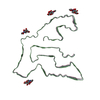
|
|---|---|
| 1 |
|
-Components
| #1: Protein | Mass: 31156.318 Da / Num. of mol.: 3 Source method: isolated from a genetically manipulated source Source: (gene. exp.) Homo sapiens (human) / Gene: TMEM106B / Production host: Homo sapiens (human) / References: UniProt: Q9NUM4#2: Sugar | ChemComp-NAG /   Type: D-saccharide, beta linking / Mass: 221.208 Da / Num. of mol.: 12 / Source method: obtained synthetically / Formula: C8H15NO6 / Feature type: SUBJECT OF INVESTIGATION Type: D-saccharide, beta linking / Mass: 221.208 Da / Num. of mol.: 12 / Source method: obtained synthetically / Formula: C8H15NO6 / Feature type: SUBJECT OF INVESTIGATIONHas ligand of interest | Y | Has protein modification | Y | |
|---|
-Experimental details
-Experiment
| Experiment | Method: ELECTRON MICROSCOPY |
|---|---|
| EM experiment | Aggregation state: FILAMENT / 3D reconstruction method: helical reconstruction |
- Sample preparation
| Component | Name: TMEM106B / Type: TISSUE / Entity ID: #1 / Source: NATURAL |
|---|---|
| Source (natural) | Organism: Homo sapiens (human) |
| Buffer solution | pH: 7.2 |
| Specimen | Conc.: 1 mg/ml / Embedding applied: NO / Shadowing applied: NO / Staining applied: NO / Vitrification applied: YES |
| Specimen support | Grid material: GRAPHENE OXIDE / Grid mesh size: 300 divisions/in. / Grid type: C-flat-1.2/1.3 |
| Vitrification | Cryogen name: ETHANE |
- Electron microscopy imaging
| Experimental equipment |  Model: Titan Krios / Image courtesy: FEI Company |
|---|---|
| Microscopy | Model: TFS KRIOS |
| Electron gun | Electron source:  FIELD EMISSION GUN / Accelerating voltage: 300 kV / Illumination mode: SPOT SCAN FIELD EMISSION GUN / Accelerating voltage: 300 kV / Illumination mode: SPOT SCAN |
| Electron lens | Mode: BRIGHT FIELD / Nominal magnification: 81000 X / Nominal defocus max: 5000 nm / Nominal defocus min: 500 nm / Cs: 2.7 mm / C2 aperture diameter: 100 µm |
| Specimen holder | Cryogen: NITROGEN |
| Image recording | Average exposure time: 1.103 sec. / Electron dose: 50.46 e/Å2 / Film or detector model: GATAN K3 (6k x 4k) |
- Processing
| EM software |
| |||||||||||||||
|---|---|---|---|---|---|---|---|---|---|---|---|---|---|---|---|---|
| CTF correction | Type: PHASE FLIPPING AND AMPLITUDE CORRECTION | |||||||||||||||
| Helical symmerty | Angular rotation/subunit: -0.43 ° / Axial rise/subunit: 4.79 Å / Axial symmetry: C1 | |||||||||||||||
| 3D reconstruction | Resolution: 3.25 Å / Resolution method: FSC 0.143 CUT-OFF / Num. of particles: 16484 / Algorithm: FOURIER SPACE / Num. of class averages: 1 / Symmetry type: HELICAL |
