 Movie
Movie Controller


+ Open data

- Basic information
| Entry | Database: PDB / ID: 7sni | |||||||||||||||||||||||||||
|---|---|---|---|---|---|---|---|---|---|---|---|---|---|---|---|---|---|---|---|---|---|---|---|---|---|---|---|---|
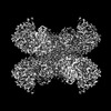
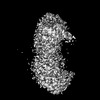
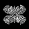
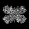
| Title | Structure of G6PD-D200N tetramer bound to NADP+ and G6P | |||||||||||||||||||||||||||
 Components Components | Glucose-6-phosphate 1-dehydrogenase | |||||||||||||||||||||||||||
 Keywords Keywords | OXIDOREDUCTASE / D200N mutant | |||||||||||||||||||||||||||
| Function / homology |  Function and homology information Function and homology informationpentose biosynthetic process / ribose phosphate biosynthetic process / response to iron(III) ion / positive regulation of calcium ion transmembrane transport via high voltage-gated calcium channel / glucose-6-phosphate dehydrogenase (NADP+) / glucose-6-phosphate dehydrogenase activity / Pentose phosphate pathway / pentose-phosphate shunt, oxidative branch / negative regulation of cell growth involved in cardiac muscle cell development / glucose 6-phosphate metabolic process ...pentose biosynthetic process / ribose phosphate biosynthetic process / response to iron(III) ion / positive regulation of calcium ion transmembrane transport via high voltage-gated calcium channel / glucose-6-phosphate dehydrogenase (NADP+) / glucose-6-phosphate dehydrogenase activity / Pentose phosphate pathway / pentose-phosphate shunt, oxidative branch / negative regulation of cell growth involved in cardiac muscle cell development / glucose 6-phosphate metabolic process / NADP+ metabolic process / pentose-phosphate shunt / D-glucose binding / NFE2L2 regulates pentose phosphate pathway genes / Oxidoreductases / cholesterol biosynthetic process / erythrocyte maturation / response to food / negative regulation of reactive oxygen species metabolic process / centriolar satellite / substantia nigra development / regulation of neuron apoptotic process / TP53 Regulates Metabolic Genes / glutathione metabolic process / lipid metabolic process / glucose metabolic process / cytoplasmic side of plasma membrane / NADP binding / cellular response to oxidative stress / response to ethanol / protein homodimerization activity / extracellular exosome / membrane / identical protein binding / cytosol / cytoplasm Similarity search - Function | |||||||||||||||||||||||||||
| Biological species |  Homo sapiens (human) Homo sapiens (human) | |||||||||||||||||||||||||||
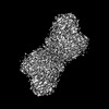
| Method | ELECTRON MICROSCOPY / single particle reconstruction / cryo EM / Resolution: 2.5 Å | |||||||||||||||||||||||||||
 Authors Authors | Wei, X. / Marmorstein, R. | |||||||||||||||||||||||||||
| Funding support |  United States, 1items United States, 1items
| |||||||||||||||||||||||||||
 Citation Citation | Journal: Proc Natl Acad Sci U S A / Year: 2022 Title: Allosteric role of a structural NADP molecule in glucose-6-phosphate dehydrogenase activity. Authors: Xuepeng Wei / Kathryn Kixmoeller / Elana Baltrusaitis / Xiaolu Yang / Ronen Marmorstein / Abstract: Human glucose-6-phosphate dehydrogenase (G6PD) is the main cellular source of NADPH, and thus plays a key role in maintaining reduced glutathione to protect cells from oxidative stress disorders such ...Human glucose-6-phosphate dehydrogenase (G6PD) is the main cellular source of NADPH, and thus plays a key role in maintaining reduced glutathione to protect cells from oxidative stress disorders such as hemolytic anemia. G6PD is a multimeric enzyme that uses the cofactors β-D-glucose 6-phosphate (G6P) and "catalytic" NADP (NADPc), as well as a "structural" NADP (NADPs) located ∼25 Å from the active site, to generate NADPH. While X-ray crystallographic and biochemical studies have revealed a role for NADPs in maintaining the catalytic activity by stabilizing the multimeric G6PD conformation, other potential roles for NADPs have not been evaluated. Here, we determined the high resolution cryo-electron microscopy structures of human wild-type G6PD in the absence of bound ligands and a catalytic G6PD-D200N mutant bound to NADPc and NADPs in the absence or presence of G6P. A comparison of these structures, together with previously reported structures, reveals that the unliganded human G6PD forms a mixture of dimers and tetramers with similar overall folds, and binding of NADPs induces a structural ordering of a C-terminal extension region and allosterically regulates G6P binding and catalysis. These studies have implications for understanding G6PD deficiencies and for therapy of G6PD-mediated disorders. | |||||||||||||||||||||||||||
| History |
|
- Structure visualization
| Structure viewer | Molecule: MolmilJmol/JSmol |
|---|
- Downloads & links
-Download
| PDBx/mmCIF format | 7sni.cif.gz | 360.5 KB | Display | PDBx/mmCIF format |
|---|---|---|---|---|
| PDB format | pdb7sni.ent.gz | 298.5 KB | Display | PDB format |
| PDBx/mmJSON format | 7sni.json.gz | Tree view | PDBx/mmJSON format | |
| Others |  Other downloads Other downloads |
-Validation report
| Arichive directory | https://data.pdbj.org/pub/pdb/validation_reports/sn/7sniftp://data.pdbj.org/pub/pdb/validation_reports/sn/7sni | HTTPS FTP |
|---|
-Related structure data
| Related structure data |  25227MC 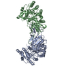 7snfC 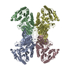 7sngC 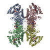 7snhC 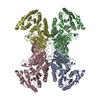 7toeC 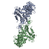 7tofC 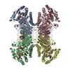 7ualC 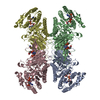 7uc2C M: map data used to model this data C: citing same article ( |
|---|---|
| Similar structure data |


-Links
 PDBj
PDBj- Assembly
| Deposited unit | 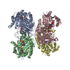
|
|---|---|
| 1 |
|
-Components
| #1: Protein | Mass: 60402.762 Da / Num. of mol.: 4 Source method: isolated from a genetically manipulated source Source: (gene. exp.) Homo sapiens (human) / Gene: G6PD / Production host:  References: UniProt: P11413, glucose-6-phosphate dehydrogenase (NADP+) #2: Chemical | ChemComp-NAP /   Mass: 743.405 Da / Num. of mol.: 8 / Source method: obtained synthetically / Formula: C21H28N7O17P3 / Feature type: SUBJECT OF INVESTIGATION Mass: 743.405 Da / Num. of mol.: 8 / Source method: obtained synthetically / Formula: C21H28N7O17P3 / Feature type: SUBJECT OF INVESTIGATION#3: Sugar | ChemComp-BG6 /   Type: D-saccharide, beta linking / Mass: 260.136 Da / Num. of mol.: 4 / Source method: obtained synthetically / Formula: C6H13O9P / Feature type: SUBJECT OF INVESTIGATION Type: D-saccharide, beta linking / Mass: 260.136 Da / Num. of mol.: 4 / Source method: obtained synthetically / Formula: C6H13O9P / Feature type: SUBJECT OF INVESTIGATION#4: Water | ChemComp-HOH / |  Mass: 18.015 Da / Num. of mol.: 233 / Source method: isolated from a natural source / Formula: H2O Mass: 18.015 Da / Num. of mol.: 233 / Source method: isolated from a natural source / Formula: H2OHas ligand of interest | Y | Has protein modification | N | |
|---|
-Experimental details
-Experiment
| Experiment | Method: ELECTRON MICROSCOPY |
|---|---|
| EM experiment | Aggregation state: PARTICLE / 3D reconstruction method: single particle reconstruction |
- Sample preparation
| Component | Name: G6PD protein / Type: ORGANELLE OR CELLULAR COMPONENT / Entity ID: #1 / Source: RECOMBINANT |
|---|---|
| Molecular weight | Value: 0.11 MDa / Experimental value: NO |
| Source (natural) | Organism: Homo sapiens (human) |
| Source (recombinant) | Organism: |
| Buffer solution | pH: 7.5 |
| Specimen | Embedding applied: NO / Shadowing applied: NO / Staining applied: NO / Vitrification applied: YES |
| Vitrification | Cryogen name: ETHANE |
- Electron microscopy imaging
| Experimental equipment |  Model: Titan Krios / Image courtesy: FEI Company |
|---|---|
| Microscopy | Model: FEI TITAN KRIOS |
| Electron gun | Electron source:  FIELD EMISSION GUN / Accelerating voltage: 300 kV / Illumination mode: FLOOD BEAM FIELD EMISSION GUN / Accelerating voltage: 300 kV / Illumination mode: FLOOD BEAM |
| Electron lens | Mode: BRIGHT FIELD / Nominal defocus max: 2000 nm / Nominal defocus min: 1000 nm |
| Image recording | Electron dose: 40 e/Å2 / Film or detector model: GATAN K3 (6k x 4k) |
- Processing
| Software | Name: PHENIX / Version: 1.19.2_4158: / Classification: refinement | ||||||||||||||||||||||||
|---|---|---|---|---|---|---|---|---|---|---|---|---|---|---|---|---|---|---|---|---|---|---|---|---|---|
| EM software | Name: PHENIX / Category: model refinement | ||||||||||||||||||||||||
| CTF correction | Type: PHASE FLIPPING AND AMPLITUDE CORRECTION | ||||||||||||||||||||||||
| 3D reconstruction | Resolution: 2.5 Å / Resolution method: FSC 0.143 CUT-OFF / Num. of particles: 67823 / Symmetry type: POINT | ||||||||||||||||||||||||
| Refine LS restraints |
|
