 Movie
Movie Controller


+ Open data

- Basic information
| Entry | Database: PDB / ID: 7scy | ||||||
|---|---|---|---|---|---|---|---|
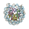
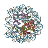
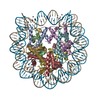
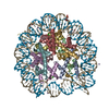
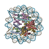
| Title | Nuc147 bound to single BRCT | ||||||
 Components Components |
| ||||||
 Keywords Keywords | DNA BINDING PROTEIN/DNA / PARP1 / BRCT / nucleosome / DNA BINDING PROTEIN-DNA complex | ||||||
| Function / homology |  Function and homology information Function and homology informationNAD+-histone H2BS6 serine ADP-ribosyltransferase activity / NAD+-histone H3S10 serine ADP-ribosyltransferase activity / NAD+-histone H2BE35 glutamate ADP-ribosyltransferase activity / positive regulation of myofibroblast differentiation / negative regulation of ATP biosynthetic process / NAD+-protein-tyrosine ADP-ribosyltransferase activity / NAD+-protein-histidine ADP-ribosyltransferase activity / regulation of base-excision repair / regulation of circadian sleep/wake cycle, non-REM sleep / mitochondrial DNA metabolic process ...NAD+-histone H2BS6 serine ADP-ribosyltransferase activity / NAD+-histone H3S10 serine ADP-ribosyltransferase activity / NAD+-histone H2BE35 glutamate ADP-ribosyltransferase activity / positive regulation of myofibroblast differentiation / negative regulation of ATP biosynthetic process / NAD+-protein-tyrosine ADP-ribosyltransferase activity / NAD+-protein-histidine ADP-ribosyltransferase activity / regulation of base-excision repair / regulation of circadian sleep/wake cycle, non-REM sleep / mitochondrial DNA metabolic process / vRNA Synthesis / carbohydrate biosynthetic process / NAD+-protein-serine ADP-ribosyltransferase activity / NAD DNA ADP-ribosyltransferase activity / negative regulation of adipose tissue development / DNA ADP-ribosylation / regulation of oxidative stress-induced neuron intrinsic apoptotic signaling pathway / ATP generation from poly-ADP-D-ribose / replication fork reversal / positive regulation of necroptotic process / signal transduction involved in regulation of gene expression / transcription regulator activator activity / response to aldosterone / HDR through MMEJ (alt-NHEJ) / positive regulation of DNA-templated transcription, elongation / NAD+ ADP-ribosyltransferase / protein auto-ADP-ribosylation / negative regulation of telomere maintenance via telomere lengthening / NAD+-protein-aspartate ADP-ribosyltransferase activity / mitochondrial DNA repair / positive regulation of intracellular estrogen receptor signaling pathway / protein poly-ADP-ribosylation / NAD+-protein-glutamate ADP-ribosyltransferase activity / negative regulation of cGAS/STING signaling pathway / positive regulation of cardiac muscle hypertrophy / NAD+-protein mono-ADP-ribosyltransferase activity / decidualization / cellular response to zinc ion / protein autoprocessing / positive regulation of mitochondrial depolarization / nuclear replication fork / R-SMAD binding / Transferases; Glycosyltransferases; Pentosyltransferases / positive regulation of SMAD protein signal transduction / negative regulation of transcription elongation by RNA polymerase II / macrophage differentiation / POLB-Dependent Long Patch Base Excision Repair / NAD+ poly-ADP-ribosyltransferase activity / negative regulation of tumor necrosis factor-mediated signaling pathway / SUMOylation of DNA damage response and repair proteins / nucleosome binding / positive regulation of double-strand break repair via homologous recombination / negative regulation of megakaryocyte differentiation / site of DNA damage / protein localization to CENP-A containing chromatin / Chromatin modifying enzymes / Replacement of protamines by nucleosomes in the male pronucleus / CENP-A containing nucleosome / protein localization to chromatin / nucleotidyltransferase activity / Packaging Of Telomere Ends / positive regulation of adipose tissue development / transforming growth factor beta receptor signaling pathway / Recognition and association of DNA glycosylase with site containing an affected purine / Cleavage of the damaged purine / epigenetic regulation of gene expression / negative regulation of innate immune response / Deposition of new CENPA-containing nucleosomes at the centromere / telomere organization / Interleukin-7 signaling / Recognition and association of DNA glycosylase with site containing an affected pyrimidine / Cleavage of the damaged pyrimidine / telomere maintenance / RNA Polymerase I Promoter Opening / Inhibition of DNA recombination at telomere / Assembly of the ORC complex at the origin of replication / nuclear estrogen receptor binding / Meiotic synapsis / SUMOylation of chromatin organization proteins / Regulation of endogenous retroelements by the Human Silencing Hub (HUSH) complex / protein modification process / DNA methylation / Condensation of Prophase Chromosomes / Chromatin modifications during the maternal to zygotic transition (MZT) / SIRT1 negatively regulates rRNA expression / HCMV Late Events / ERCC6 (CSB) and EHMT2 (G9a) positively regulate rRNA expression / response to gamma radiation / PRC2 methylates histones and DNA / Regulation of endogenous retroelements by KRAB-ZFP proteins / Defective pyroptosis / innate immune response in mucosa / HDACs deacetylate histones / mitochondrion organization / Regulation of endogenous retroelements by Piwi-interacting RNAs (piRNAs) / RNA Polymerase I Promoter Escape / Nonhomologous End-Joining (NHEJ) / lipopolysaccharide binding / Downregulation of SMAD2/3:SMAD4 transcriptional activity / Transcriptional regulation by small RNAs Similarity search - Function | ||||||
| Biological species |  Homo sapiens (human) Homo sapiens (human) | ||||||
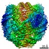
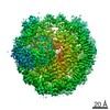
| Method | ELECTRON MICROSCOPY / single particle reconstruction / cryo EM / Resolution: 4.1 Å | ||||||
 Authors Authors | Muthurajan, U.M. / Rudolph, J.R. | ||||||
| Funding support |  United States, 1items United States, 1items
| ||||||
 Citation Citation | Journal: Mol Cell / Year: 2021 Title: The BRCT domain of PARP1 binds intact DNA and mediates intrastrand transfer. Authors: Johannes Rudolph / Uma M Muthurajan / Megan Palacio / Jyothi Mahadevan / Genevieve Roberts / Annette H Erbse / Pamela N Dyer / Karolin Luger / Abstract: PARP1 is a key player in the response to DNA damage and is the target of clinical inhibitors for the treatment of cancers. Binding of PARP1 to damaged DNA leads to activation wherein PARP1 uses NAD ...PARP1 is a key player in the response to DNA damage and is the target of clinical inhibitors for the treatment of cancers. Binding of PARP1 to damaged DNA leads to activation wherein PARP1 uses NAD to add chains of poly(ADP-ribose) onto itself and other nuclear proteins. PARP1 also binds abundantly to intact DNA and chromatin, where it remains enzymatically inactive. We show that intact DNA makes contacts with the PARP1 BRCT domain, which was not previously recognized as a DNA-binding domain. This binding mode does not result in the concomitant reorganization and activation of the catalytic domain. We visualize the BRCT domain bound to nucleosomal DNA by cryogenic electron microscopy and identify a key motif conserved from ancestral BRCT domains for binding phosphates on DNA and phospho-peptides. Finally, we demonstrate that the DNA-binding properties of the BRCT domain contribute to the "monkey-bar mechanism" that mediates DNA transfer of PARP1. | ||||||
| History |
|
- Structure visualization
| Movie |
Movie viewer |
|---|---|
| Structure viewer | Molecule: MolmilJmol/JSmol |
- Downloads & links
-Download
| PDBx/mmCIF format | 7scy.cif.gz | 528.4 KB | Display | PDBx/mmCIF format |
|---|---|---|---|---|
| PDB format | pdb7scy.ent.gz | 416.8 KB | Display | PDB format |
| PDBx/mmJSON format | 7scy.json.gz | Tree view | PDBx/mmJSON format | |
| Others |  Other downloads Other downloads |
-Validation report
| Arichive directory | https://data.pdbj.org/pub/pdb/validation_reports/sc/7scyftp://data.pdbj.org/pub/pdb/validation_reports/sc/7scy | HTTPS FTP |
|---|
-Related structure data
| Related structure data |  25042MC 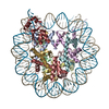 7sczC M: map data used to model this data C: citing same article ( |
|---|---|
| Similar structure data |


-Links
 PDBj
PDBj


- Assembly
| Deposited unit | 
|
|---|---|
| 1 |
|
-Components
-DNA chain , 2 types, 2 molecules IJ
| #1: DNA chain | Mass: 45610.043 Da / Num. of mol.: 1 / Source method: obtained synthetically / Source: (synth.) Homo sapiens (human) |
|---|---|
| #2: DNA chain | Mass: 45138.770 Da / Num. of mol.: 1 / Source method: obtained synthetically / Source: (synth.) Homo sapiens (human) |
-Protein , 5 types, 9 molecules AEBFCGDHK
| #3: Protein | Mass: 15719.445 Da / Num. of mol.: 2 Source method: isolated from a genetically manipulated source Source: (gene. exp.) Homo sapiens (human)Gene: H3C1, H3FA, HIST1H3A, H3C2, H3FL, HIST1H3B, H3C3, H3FC HIST1H3C, H3C4, H3FB, HIST1H3D, H3C6, H3FD, HIST1H3E, H3C7, H3FI, HIST1H3F, H3C8, H3FH, HIST1H3G, H3C10, H3FK, HIST1H3H, H3C11, H3FF, ...Gene: H3C1, H3FA, HIST1H3A, H3C2, H3FL, HIST1H3B, H3C3, H3FC HIST1H3C, H3C4, H3FB, HIST1H3D, H3C6, H3FD, HIST1H3E, H3C7, H3FI, HIST1H3F, H3C8, H3FH, HIST1H3G, H3C10, H3FK, HIST1H3H, H3C11, H3FF, HIST1H3I, H3C12, H3FJ, HIST1H3J Production host:  #4: Protein | Mass: 11676.703 Da / Num. of mol.: 2 Source method: isolated from a genetically manipulated source Source: (gene. exp.) Homo sapiens (human)Gene: H4C1, H4/A, H4FA, HIST1H4A, H4C2, H4/I, H4FI, HIST1H4B, H4C3, H4/G, H4FG, HIST1H4C, H4C4, H4/B, H4FB, HIST1H4D, H4C5, H4/J, H4FJ, HIST1H4E, H4C6, H4/C, H4FC, HIST1H4F, H4C8, H4/H, H4FH, ...Gene: H4C1, H4/A, H4FA, HIST1H4A, H4C2, H4/I, H4FI, HIST1H4B, H4C3, H4/G, H4FG, HIST1H4C, H4C4, H4/B, H4FB, HIST1H4D, H4C5, H4/J, H4FJ, HIST1H4E, H4C6, H4/C, H4FC, HIST1H4F, H4C8, H4/H, H4FH, HIST1H4H, H4C9, H4/M, H4FM, HIST1H4I, H4C11, H4/E, H4FE, HIST1H4J, H4C12, H4/D, H4FD, HIST1H4K, H4C13, H4/K, H4FK, HIST1H4L, H4C14, H4/N, H4F2, H4FN, HIST2H4, HIST2H4A, H4C15, H4/O, H4FO, HIST2H4B, H4-16, HIST4H4 Production host: #5: Protein | Mass: 14447.825 Da / Num. of mol.: 2 Source method: isolated from a genetically manipulated source Source: (gene. exp.) Homo sapiens (human) / Gene: HIST1H2AB, HIST1H2AE, hCG_1640984, hCG_1787383 / Production host: #6: Protein | Mass: 14217.516 Da / Num. of mol.: 2 Source method: isolated from a genetically manipulated source Source: (gene. exp.) Homo sapiens (human) / Gene: HIST1H2BJ, H2BFR / Production host: #7: Protein | | Mass: 14337.548 Da / Num. of mol.: 1 Source method: isolated from a genetically manipulated source Source: (gene. exp.) Homo sapiens (human) / Gene: PARP1, ADPRT, PPOL / Production host: References: UniProt: P09874, NAD+ ADP-ribosyltransferase, Transferases; Glycosyltransferases; Pentosyltransferases |
|---|
-Experimental details
-Experiment
| Experiment | Method: ELECTRON MICROSCOPY |
|---|---|
| EM experiment | Aggregation state: PARTICLE / 3D reconstruction method: single particle reconstruction |
- Sample preparation
| Component | Name: Nuc147-PARP1-BRCT / Type: COMPLEX / Entity ID: all / Source: MULTIPLE SOURCES |
|---|---|
| Molecular weight | Value: 0.214 MDa / Experimental value: NO |
| Source (natural) | Organism: Homo sapiens (human) |
| Source (recombinant) | Organism: |
| Buffer solution | pH: 7.5 |
| Specimen | Embedding applied: NO / Shadowing applied: NO / Staining applied: NO / Vitrification applied: YES |
| Vitrification | Cryogen name: ETHANE |
- Electron microscopy imaging
| Experimental equipment |  Model: Titan Krios / Image courtesy: FEI Company |
|---|---|
| Microscopy | Model: FEI TITAN KRIOS |
| Electron gun | Electron source:  FIELD EMISSION GUN / Accelerating voltage: 300 kV / Illumination mode: OTHER FIELD EMISSION GUN / Accelerating voltage: 300 kV / Illumination mode: OTHER |
| Electron lens | Mode: OTHER |
| Image recording | Electron dose: 60 e/Å2 / Film or detector model: GATAN K3 (6k x 4k) |
- Processing
| CTF correction | Type: NONE |
|---|---|
| 3D reconstruction | Resolution: 4.1 Å / Resolution method: FSC 0.143 CUT-OFF / Num. of particles: 16495 / Symmetry type: POINT |
