- PDB-7oto: The structure of MutS bound to two molecules of AMPPNP -
+
データを開く
IDまたはキーワード:
読み込み中...
-
基本情報
登録情報
データベース: PDB / ID: 7oto
タイトル
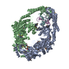
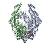
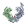
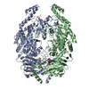
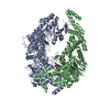
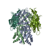
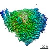
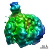
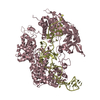
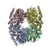
The structure of MutS bound to two molecules of AMPPNP
要素
DNA mismatch repair protein MutS
キーワード
DNA BINDING PROTEIN / DNA mismatch repair protein
機能・相同性
機能・相同性情報
adenine/cytosine mispair binding / MutS complex / mismatch repair complex / regulation of DNA recombination / mismatched DNA binding / DNA binding, bending / ATP-dependent DNA damage sensor activity / mismatch repair / ADP binding / damaged DNA binding ...adenine/cytosine mispair binding / MutS complex / mismatch repair complex / regulation of DNA recombination / mismatched DNA binding / DNA binding, bending / ATP-dependent DNA damage sensor activity / mismatch repair / ADP binding / damaged DNA binding / DNA damage response / ATP hydrolysis activity / ATP binding / identical protein binding / cytosol 類似検索 - 分子機能
DNA mismatch repair protein MutS / DNA mismatch repair protein MutS/MSH / DNA mismatch repair protein MutS-like, N-terminal / DNA mismatch repair protein MutS, connector domain / DNA mismatch repair protein MutS, clamp / DNA mismatch repair protein MutS, N-terminal / MutS, connector domain superfamily / MutS domain I / MutS domain II / MutS family domain IV ...DNA mismatch repair protein MutS / DNA mismatch repair protein MutS/MSH / DNA mismatch repair protein MutS-like, N-terminal / DNA mismatch repair protein MutS, connector domain / DNA mismatch repair protein MutS, clamp / DNA mismatch repair protein MutS, N-terminal / MutS, connector domain superfamily / MutS domain I / MutS domain II / MutS family domain IV / MutS domain III / DNA mismatch repair MutS family / DNA mismatch repair protein MutS, C-terminal / DNA mismatch repair protein MutS, core / DNA mismatch repair protein MutS, core domain superfamily / MutS domain V / DNA mismatch repair proteins mutS family signature. / DNA-binding domain of DNA mismatch repair MUTS family / ATPase domain of DNA mismatch repair MUTS family / P-loop containing nucleoside triphosphate hydrolase 類似検索 - ドメイン・相同性
PHOSPHOAMINOPHOSPHONIC ACID-ADENYLATE ESTER / DNA mismatch repair protein MutS 類似検索 - 構成要素
ジャーナル: Nat Struct Mol Biol / 年: 2022 タイトル: Cryogenic electron microscopy structures reveal how ATP and DNA binding in MutS coordinates sequential steps of DNA mismatch repair. 著者: Alessandro Borsellini / Vladislav Kunetsky / Peter Friedhoff / Meindert H Lamers / 要旨: DNA mismatch repair detects and corrects mismatches introduced during DNA replication. The protein MutS scans for mismatches and coordinates the repair cascade. During this process, MutS undergoes ...DNA mismatch repair detects and corrects mismatches introduced during DNA replication. The protein MutS scans for mismatches and coordinates the repair cascade. During this process, MutS undergoes multiple conformational changes in response to ATP binding, hydrolysis and release, but how ATP induces the various MutS conformations is incompletely understood. Here we present four cryogenic electron microscopy structures of Escherichia coli MutS at sequential stages of the ATP hydrolysis cycle that reveal how ATP binding and hydrolysis induce closing and opening of the MutS dimer, respectively. Biophysical analysis demonstrates how DNA binding modulates the ATPase cycle by prevention of hydrolysis during scanning and mismatch binding, while preventing ADP release in the sliding clamp state. Nucleotide release is achieved when MutS encounters single-stranded DNA that is produced during removal of the daughter strand. The combination of ATP binding and hydrolysis and its modulation by DNA enables MutS to adopt the different conformations needed to coordinate the sequential steps of the mismatch repair cascade.
分子量: 506.196 Da / 分子数: 2 / 由来タイプ: 合成 / 式: C10H17N6O12P3 / タイプ: SUBJECT OF INVESTIGATION コメント: AMP-PNP, エネルギー貯蔵分子類似体*YM
研究の焦点であるリガンドがあるか
Y
-
実験情報
-
実験
実験
手法: 電子顕微鏡法
EM実験
試料の集合状態: PARTICLE / 3次元再構成法: 単粒子再構成法
-
試料調製
構成要素
名称: DNA mismatch repair protein MutS / タイプ: ORGANELLE OR CELLULAR COMPONENT / 詳細: MutS bound to two molecules of AMPPNP / Entity ID: #1 / 由来: RECOMBINANT
 ムービー
ムービー コントローラー


 データを開く
 基本情報
基本情報 要素
要素 キーワード
キーワード 機能・相同性情報
 データ登録者
 引用
引用

 構造の表示
構造の表示 ダウンロードとリンク
 その他のダウンロード
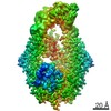


 PDBj
PDBj


 集合体
集合体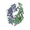

 分子量: 24.305 Da / 分子数: 2 / 由来タイプ: 合成 / 式: Mg
 分子量: 506.196 Da / 分子数: 2 / 由来タイプ: 合成 / 式: C10H17N6O12P3 / タイプ: SUBJECT OF INVESTIGATION
 試料調製
試料調製 電子顕微鏡撮影
 FIELD EMISSION GUN / 加速電圧: 300 kV / 照射モード: FLOOD BEAM
 解析
解析