 Movie
Movie Controller

[English] 日本語
 Yorodumi
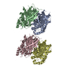
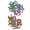
- PDB-7om8: Beta2 appendage domain of AP2 bound to terminal domains beneath t... -
+ Open data

- Basic information
| Entry | Database: PDB / ID: 7om8 | ||||||||||||
|---|---|---|---|---|---|---|---|---|---|---|---|---|---|
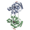
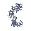
| Title | Beta2 appendage domain of AP2 bound to terminal domains beneath the hub of the 28 triskelia mini clathrin coat complex, class 15 | ||||||||||||
 Components Components |
| ||||||||||||
 Keywords Keywords | ENDOCYTOSIS / Clathrin / clathrin adaptor / AP2 | ||||||||||||
| Function / homology |  Function and homology information Function and homology informationclathrin coat of trans-Golgi network vesicle / Nef Mediated CD8 Down-regulation / clathrin light chain binding / WNT5A-dependent internalization of FZD2, FZD5 and ROR2 / clathrin complex / Trafficking of GluR2-containing AMPA receptors / WNT5A-dependent internalization of FZD4 / clathrin adaptor complex / extrinsic component of presynaptic endocytic zone membrane / clathrin coat of coated pit ...clathrin coat of trans-Golgi network vesicle / Nef Mediated CD8 Down-regulation / clathrin light chain binding / WNT5A-dependent internalization of FZD2, FZD5 and ROR2 / clathrin complex / Trafficking of GluR2-containing AMPA receptors / WNT5A-dependent internalization of FZD4 / clathrin adaptor complex / extrinsic component of presynaptic endocytic zone membrane / clathrin coat of coated pit / postsynaptic endocytic zone / postsynaptic neurotransmitter receptor internalization / AP-2 adaptor complex / Retrograde neurotrophin signalling / clathrin-coated endocytic vesicle / clathrin coat assembly / LDL clearance / clathrin-dependent endocytosis / signal sequence receptor activity / Nef Mediated CD4 Down-regulation / coronary vasculature development / positive regulation of protein localization to membrane / endolysosome membrane / neurotransmitter secretion / ventricular septum development / aorta development / clathrin binding / Recycling pathway of L1 / EPH-ephrin mediated repulsion of cells / positive regulation of endocytosis / synaptic vesicle endocytosis / vesicle-mediated transport / MHC class II antigen presentation / receptor-mediated endocytosis / VLDLR internalisation and degradation / kidney development / clathrin-coated endocytic vesicle membrane / intracellular protein transport / spindle / cytoplasmic side of plasma membrane / endocytic vesicle membrane / synaptic vesicle / Cargo recognition for clathrin-mediated endocytosis / mitotic cell cycle / Clathrin-mediated endocytosis / Potential therapeutics for SARS / protein-containing complex binding / structural molecule activity / glutamatergic synapse / membrane / plasma membrane / cytosol Similarity search - Function | ||||||||||||
| Biological species |  Homo sapiens (human) Homo sapiens (human) | ||||||||||||
| Method | ELECTRON MICROSCOPY / single particle reconstruction / cryo EM / Resolution: 10.5 Å | ||||||||||||
 Authors Authors | Smith, S.M. / Smith, C.J. | ||||||||||||
| Funding support |  United Kingdom, 1items United Kingdom, 1items
| ||||||||||||
 Citation Citation | Journal: EMBO J / Year: 2021 Title: Multi-modal adaptor-clathrin contacts drive coated vesicle assembly. Authors: Sarah M Smith / Gabrielle Larocque / Katherine M Wood / Kyle L Morris / Alan M Roseman / Richard B Sessions / Stephen J Royle / Corinne J Smith / Abstract: Clathrin-coated pits are formed by the recognition of membrane and cargo by the AP2 complex and the subsequent recruitment of clathrin triskelia. A role for AP2 in coated-pit assembly beyond initial ...Clathrin-coated pits are formed by the recognition of membrane and cargo by the AP2 complex and the subsequent recruitment of clathrin triskelia. A role for AP2 in coated-pit assembly beyond initial clathrin recruitment has not been explored. Clathrin binds the β2 subunit of AP2, and several binding sites have been identified, but our structural knowledge of these interactions is incomplete and their functional importance during endocytosis is unclear. Here, we analysed the cryo-EM structure of clathrin cages assembled in the presence of β2 hinge-appendage (β2HA). We find that the β2-appendage binds in at least two positions in the cage, demonstrating that multi-modal binding is a fundamental property of clathrin-AP2 interactions. In one position, β2-appendage cross-links two adjacent terminal domains from different triskelia. Functional analysis of β2HA-clathrin interactions reveals that endocytosis requires two clathrin interaction sites: a clathrin-box motif on the hinge and the "sandwich site" on the appendage. We propose that β2-appendage binding to more than one triskelion is a key feature of the system and likely explains why assembly is driven by AP2. | ||||||||||||
| History |
|
- Structure visualization
| Movie |
Movie viewer |
|---|---|
| Structure viewer | Molecule: MolmilJmol/JSmol |
- Downloads & links
-Download
| PDBx/mmCIF format | 7om8.cif.gz | 192.2 KB | Display | PDBx/mmCIF format |
|---|---|---|---|---|
| PDB format | pdb7om8.ent.gz | 147.3 KB | Display | PDB format |
| PDBx/mmJSON format | 7om8.json.gz | Tree view | PDBx/mmJSON format | |
| Others |  Other downloads Other downloads |
-Validation report
| Arichive directory | https://data.pdbj.org/pub/pdb/validation_reports/om/7om8ftp://data.pdbj.org/pub/pdb/validation_reports/om/7om8 | HTTPS FTP |
|---|
-Related structure data
| Related structure data |  12984MC M: map data used to model this data C: citing same article ( |
|---|---|
| Similar structure data | |
| EM raw data | EMPIAR-10783 (Title: Multi-modal adaptor-clathrin contacts drive coated vesicle assembly Data size: 25.0 Data #1: Hub subparticles of the 28 mini coat, class 15 [picked particles - single frame - processed]) |


-Links
 PDBj
PDBj


- Assembly
| Deposited unit | 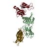
|
|---|---|
| 1 |
|
-Components
| #1: Antibody | Mass: 33535.574 Da / Num. of mol.: 2 / Source method: isolated from a natural source / Source: (natural) #2: Protein | | Mass: 26429.457 Da / Num. of mol.: 1 Source method: isolated from a genetically manipulated source Source: (gene. exp.) Homo sapiens (human) / Gene: AP2B1, ADTB2, CLAPB1 / Production host:  |
|---|
-Experimental details
-Experiment
| Experiment | Method: ELECTRON MICROSCOPY |
|---|---|
| EM experiment | Aggregation state: PARTICLE / 3D reconstruction method: single particle reconstruction |
- Sample preparation
| Component |
| ||||||||||||||||||||||||
|---|---|---|---|---|---|---|---|---|---|---|---|---|---|---|---|---|---|---|---|---|---|---|---|---|---|
| Molecular weight | Experimental value: NO | ||||||||||||||||||||||||
| Source (natural) |
| ||||||||||||||||||||||||
| Source (recombinant) | Organism: | ||||||||||||||||||||||||
| Buffer solution | pH: 6.4 | ||||||||||||||||||||||||
| Specimen | Embedding applied: NO / Shadowing applied: NO / Staining applied: NO / Vitrification applied: YES | ||||||||||||||||||||||||
| Vitrification | Instrument: LEICA EM GP / Cryogen name: ETHANE-PROPANE / Humidity: 90 % / Chamber temperature: 277.15 K Details: 3 uL of sample applied to a grid and blotted for 4.5 s before plunging |
- Electron microscopy imaging
| Experimental equipment |  Model: Titan Krios / Image courtesy: FEI Company |
|---|---|
| Microscopy | Model: FEI TITAN KRIOS |
| Electron gun | Electron source:  FIELD EMISSION GUN / Accelerating voltage: 300 kV / Illumination mode: FLOOD BEAM FIELD EMISSION GUN / Accelerating voltage: 300 kV / Illumination mode: FLOOD BEAM |
| Electron lens | Mode: BRIGHT FIELD / Nominal magnification: 59000 X / Nominal defocus max: 2000 nm / Nominal defocus min: 1100 nm / Cs: 2.7 mm |
| Specimen holder | Specimen holder model: FEI TITAN KRIOS AUTOGRID HOLDER |
| Image recording | Electron dose: 64 e/Å2 / Film or detector model: FEI FALCON III (4k x 4k) |
- Processing
| EM software | Name: RELION / Version: 3.0.5 / Category: CTF correction |
|---|---|
| CTF correction | Type: PHASE FLIPPING AND AMPLITUDE CORRECTION |
| Symmetry | Point symmetry: C1 (asymmetric) |
| 3D reconstruction | Resolution: 10.5 Å / Resolution method: FSC 0.143 CUT-OFF / Num. of particles: 11676 / Symmetry type: POINT |
| Atomic model building | Details: This structural model has been updated to include only clathrin terminal domain beta propeller residues that are close to the interface with the beta2 appendage domain. |
| Refinement | Highest resolution: 10.5 Å |
