 Movie
Movie Controller

[English] 日本語
 Yorodumi
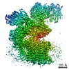
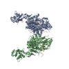
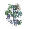
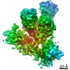
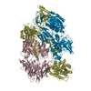
- PDB-7oci: Cryo-EM structure of yeast Ost6p containing oligosaccharyltransfe... -
+ Open data

- Basic information
| Entry | Database: PDB / ID: 7oci | |||||||||||||||||||||||||||
|---|---|---|---|---|---|---|---|---|---|---|---|---|---|---|---|---|---|---|---|---|---|---|---|---|---|---|---|---|
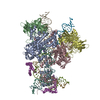
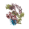
| Title | Cryo-EM structure of yeast Ost6p containing oligosaccharyltransferase complex | |||||||||||||||||||||||||||
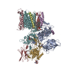
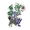
 Components Components | (Dolichyl-diphosphooligosaccharide--protein glycosyltransferase subunit ...) x 9 | |||||||||||||||||||||||||||
 Keywords Keywords | MEMBRANE PROTEIN / N-glycosylation / Yeast / Complex / Endoplasmic reticulum | |||||||||||||||||||||||||||
| Function / homology |  Function and homology information Function and homology informationMiscellaneous transport and binding events / dolichol-linked oligosaccharide biosynthetic process / dolichyl-diphosphooligosaccharide-protein glycotransferase / dolichyl-diphosphooligosaccharide-protein glycotransferase activity / oligosaccharyltransferase complex / : / protein N-linked glycosylation / glycosyltransferase activity / protein-disulfide reductase activity / Neutrophil degranulation ...Miscellaneous transport and binding events / dolichol-linked oligosaccharide biosynthetic process / dolichyl-diphosphooligosaccharide-protein glycotransferase / dolichyl-diphosphooligosaccharide-protein glycotransferase activity / oligosaccharyltransferase complex / : / protein N-linked glycosylation / glycosyltransferase activity / protein-disulfide reductase activity / Neutrophil degranulation / post-translational protein modification / nuclear envelope / protein-macromolecule adaptor activity / endoplasmic reticulum membrane / structural molecule activity / endoplasmic reticulum / mitochondrion / metal ion binding Similarity search - Function | |||||||||||||||||||||||||||
| Biological species |  | |||||||||||||||||||||||||||
| Method | ELECTRON MICROSCOPY / single particle reconstruction / cryo EM / Resolution: 3.46 Å | |||||||||||||||||||||||||||
 Authors Authors | Wild, R. / Neuhaus, J.D. / Eyring, J. / Irobalieva, R.N. / Kowal, J. / Lin, C.W. / Locher, K.P. / Aebi, M. | |||||||||||||||||||||||||||
| Funding support |  Switzerland, 1items Switzerland, 1items
| |||||||||||||||||||||||||||
 Citation Citation | Journal: Glycobiology / Year: 2021 Title: Functional analysis of Ost3p and Ost6p containing yeast oligosaccharyltransferases. Abstract: The oligosaccharyltransferase (OST) is the central enzyme in the N-glycosylation pathway. It transfers a defined oligosaccharide from a lipid-linker onto the asparagine side chain of proteins. The ...The oligosaccharyltransferase (OST) is the central enzyme in the N-glycosylation pathway. It transfers a defined oligosaccharide from a lipid-linker onto the asparagine side chain of proteins. The yeast OST consists of eight subunits and exists in two catalytically distinct isoforms that differ in one subunit, Ost3p or Ost6p. The cryo-electron microscopy structure of the Ost6p containing complex was found to be highly similar to the Ost3p containing OST. OST enzymes with altered Ost3p/Ost6p subunits were generated and functionally analyzed. The three C-terminal transmembrane helices were responsible for the higher turnover-rate of the Ost3p vs. the Ost6p containing enzyme in vitro and the more severe hypoglycosylation in Ost3p lacking strains in vivo. Glycosylation of specific OST target sites required the N-terminal thioredoxin domain of Ost3p or Ost6p. This Ost3p/Ost6p dependence was glycosylation site but not protein specific. We concluded that the Ost3p/Ost6p subunits modulate the catalytic activity of OST and provide additional specificity for OST substrate recognition. | |||||||||||||||||||||||||||
| History |
|
- Structure visualization
| Movie |
Movie viewer |
|---|---|
| Structure viewer | Molecule: MolmilJmol/JSmol |
- Downloads & links
-Download
| PDBx/mmCIF format | 7oci.cif.gz | 403.5 KB | Display | PDBx/mmCIF format |
|---|---|---|---|---|
| PDB format | pdb7oci.ent.gz | 315.1 KB | Display | PDB format |
| PDBx/mmJSON format | 7oci.json.gz | Tree view | PDBx/mmJSON format | |
| Others |  Other downloads Other downloads |
-Validation report
| Arichive directory | https://data.pdbj.org/pub/pdb/validation_reports/oc/7ociftp://data.pdbj.org/pub/pdb/validation_reports/oc/7oci | HTTPS FTP |
|---|
-Related structure data
| Related structure data | 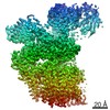 12808MC M: map data used to model this data C: citing same article ( |
|---|---|
| Similar structure data |

-Links
 PDBj
PDBj


- Assembly
| Deposited unit | 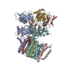
|
|---|---|
| 1 |
|
-Components
-Dolichyl-diphosphooligosaccharide--protein glycosyltransferase subunit ... , 9 types, 9 molecules ABCDEFGHI
| #1: Protein | Mass: 54116.477 Da / Num. of mol.: 1 / Source method: isolated from a natural source / Source: (natural) References: UniProt: P41543, dolichyl-diphosphooligosaccharide-protein glycotransferase |
|---|---|
| #2: Protein | Mass: 14712.531 Da / Num. of mol.: 1 / Source method: isolated from a natural source / Source: (natural) References: UniProt: P46964, dolichyl-diphosphooligosaccharide-protein glycotransferase |
| #3: Protein | Mass: 37921.344 Da / Num. of mol.: 1 / Source method: isolated from a natural source / Source: (natural) |
| #4: Protein/peptide | Mass: 3986.696 Da / Num. of mol.: 1 / Source method: isolated from a natural source / Source: (natural) References: UniProt: Q99380, dolichyl-diphosphooligosaccharide-protein glycotransferase |
| #5: Protein | Mass: 9525.090 Da / Num. of mol.: 1 / Source method: isolated from a natural source / Source: (natural) References: UniProt: Q92316, dolichyl-diphosphooligosaccharide-protein glycotransferase |
| #6: Protein | Mass: 81604.539 Da / Num. of mol.: 1 / Source method: isolated from a natural source / Source: (natural) References: UniProt: P39007, dolichyl-diphosphooligosaccharide-protein glycotransferase |
| #7: Protein | Mass: 49444.438 Da / Num. of mol.: 1 / Source method: isolated from a natural source / Source: (natural) References: UniProt: P33767, dolichyl-diphosphooligosaccharide-protein glycotransferase |
| #8: Protein | Mass: 31682.832 Da / Num. of mol.: 1 / Source method: isolated from a natural source / Source: (natural) References: UniProt: Q02795, dolichyl-diphosphooligosaccharide-protein glycotransferase |
| #9: Protein/peptide | Mass: 2060.531 Da / Num. of mol.: 1 / Source method: isolated from a natural source / Details: alpha-helix modelled as poly-UNK / Source: (natural) References: dolichyl-diphosphooligosaccharide-protein glycotransferase |
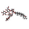
-Sugars , 3 types, 5 molecules 
| #10: Polysaccharide | 2-acetamido-2-deoxy-beta-D-glucopyranose-(1-4)-2-acetamido-2-deoxy-beta-D-glucopyranose |
|---|---|
| #11: Polysaccharide | alpha-D-mannopyranose-(1-2)-alpha-D-mannopyranose-(1-2)-alpha-D-mannopyranose-(1-3)-[alpha-D- ...alpha-D-mannopyranose-(1-2)-alpha-D-mannopyranose-(1-2)-alpha-D-mannopyranose-(1-3)-[alpha-D-mannopyranose-(1-6)]beta-D-mannopyranose-(1-4)-2-acetamido-2-deoxy-beta-D-glucopyranose-(1-4)-2-acetamido-2-deoxy-beta-D-glucopyranose Source method: isolated from a genetically manipulated source |
| #13: Sugar |  Type: D-saccharide, beta linking / Mass: 221.208 Da / Num. of mol.: 3 / Source method: obtained synthetically / Formula: C8H15NO6 Type: D-saccharide, beta linking / Mass: 221.208 Da / Num. of mol.: 3 / Source method: obtained synthetically / Formula: C8H15NO6 |
-Non-polymers , 4 types, 10 molecules 

| #12: Chemical | ChemComp-PEE /  Mass: 744.034 Da / Num. of mol.: 6 / Source method: obtained synthetically / Formula: C41H78NO8P / Comment: DOPE, phospholipid*YM Mass: 744.034 Da / Num. of mol.: 6 / Source method: obtained synthetically / Formula: C41H78NO8P / Comment: DOPE, phospholipid*YM#14: Chemical |  Mass: 1229.312 Da / Num. of mol.: 2 / Source method: obtained synthetically / Formula: C56H92O29 / Comment: detergent*YM Mass: 1229.312 Da / Num. of mol.: 2 / Source method: obtained synthetically / Formula: C56H92O29 / Comment: detergent*YM#15: Chemical | ChemComp-MG / |  Mass: 24.305 Da / Num. of mol.: 1 / Source method: obtained synthetically / Formula: Mg Mass: 24.305 Da / Num. of mol.: 1 / Source method: obtained synthetically / Formula: Mg#16: Chemical | ChemComp-V8K / |  Mass: 847.282 Da / Num. of mol.: 1 / Source method: obtained synthetically / Formula: C55H91O4P Mass: 847.282 Da / Num. of mol.: 1 / Source method: obtained synthetically / Formula: C55H91O4P |
|---|
-Details
| Has ligand of interest | N |
|---|---|
| Has protein modification | Y |
-Experimental details
-Experiment
| Experiment | Method: ELECTRON MICROSCOPY |
|---|---|
| EM experiment | Aggregation state: PARTICLE / 3D reconstruction method: single particle reconstruction |
- Sample preparation
| Component | Name: Ost6p containing yeast oligosaccharyltransferase complex Type: COMPLEX / Entity ID: #2-#9 / Source: NATURAL |
|---|---|
| Source (natural) | Organism: |
| Buffer solution | pH: 7.5 |
| Specimen | Conc.: 5 mg/ml / Embedding applied: NO / Shadowing applied: NO / Staining applied: NO / Vitrification applied: YES / Details: This sample was monodisperse |
| Specimen support | Grid material: COPPER / Grid mesh size: 300 divisions/in. / Grid type: Quantifoil R1.2/1.3 |
| Vitrification | Cryogen name: ETHANE-PROPANE |
- Electron microscopy imaging
| Experimental equipment |  Model: Titan Krios / Image courtesy: FEI Company |
|---|---|
| Microscopy | Model: FEI TITAN KRIOS |
| Electron gun | Electron source:  FIELD EMISSION GUN / Accelerating voltage: 300 kV / Illumination mode: FLOOD BEAM FIELD EMISSION GUN / Accelerating voltage: 300 kV / Illumination mode: FLOOD BEAM |
| Electron lens | Mode: BRIGHT FIELD / C2 aperture diameter: 2.7 µm |
| Specimen holder | Specimen holder model: FEI TITAN KRIOS AUTOGRID HOLDER |
| Image recording | Electron dose: 67 e/Å2 / Film or detector model: GATAN K2 SUMMIT (4k x 4k) |
- Processing
| Software | Name: PHENIX / Version: 1.19.1_4122: / Classification: refinement | ||||||||||||||||||||||||
|---|---|---|---|---|---|---|---|---|---|---|---|---|---|---|---|---|---|---|---|---|---|---|---|---|---|
| EM software |
| ||||||||||||||||||||||||
| CTF correction | Type: PHASE FLIPPING AND AMPLITUDE CORRECTION | ||||||||||||||||||||||||
| 3D reconstruction | Resolution: 3.46 Å / Resolution method: FSC 0.143 CUT-OFF / Num. of particles: 220536 / Symmetry type: POINT | ||||||||||||||||||||||||
| Atomic model building | Protocol: RIGID BODY FIT | ||||||||||||||||||||||||
| Atomic model building | PDB-ID: 6EZN Accession code: 6EZN / Source name: PDB / Type: experimental model | ||||||||||||||||||||||||
| Refine LS restraints |
|

