ムービー
ムービー コントローラー


+ データを開く

- 基本情報
基本情報
| 登録情報 | データベース: PDB / ID: 7nzm | ||||||
|---|---|---|---|---|---|---|---|
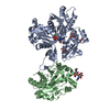
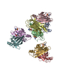
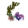
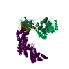
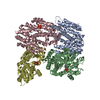
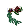
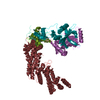
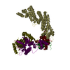
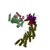
| タイトル | Cryo-EM structure of pre-dephosphorylation complex of phosphorylated eIF2alpha with trapped holophosphatase (PP1A_D64A/PPP1R15A/G-actin/DNase I) | ||||||
 要素 要素 |
| ||||||
 キーワード キーワード | HYDROLASE / holophosphatase / PP1 / PPP1R15A / phosphorylated eIF2alpha | ||||||
| 機能・相同性 |  機能・相同性情報 機能・相同性情報fatty acid derivative binding / positive regulation of translational initiation in response to stress / positive regulation of endoplasmic reticulum stress-induced eIF2 alpha dephosphorylation / regulation of translational initiation by eIF2 alpha dephosphorylation / regulation of neutrophil mediated cytotoxicity / zymogen granule / regulation of acute inflammatory response / positive regulation of peptidyl-serine dephosphorylation / regulation of translation in response to endoplasmic reticulum stress / translation initiation ternary complex ...fatty acid derivative binding / positive regulation of translational initiation in response to stress / positive regulation of endoplasmic reticulum stress-induced eIF2 alpha dephosphorylation / regulation of translational initiation by eIF2 alpha dephosphorylation / regulation of neutrophil mediated cytotoxicity / zymogen granule / regulation of acute inflammatory response / positive regulation of peptidyl-serine dephosphorylation / regulation of translation in response to endoplasmic reticulum stress / translation initiation ternary complex / protein phosphatase type 1 complex / glial limiting end-foot / response to kainic acid / Cellular response to mitochondrial stress / deoxyribonuclease I / response to manganese-induced endoplasmic reticulum stress / positive regulation of type B pancreatic cell apoptotic process / negative regulation of translational initiation in response to stress / Response of EIF2AK1 (HRI) to heme deficiency / PTW/PP1 phosphatase complex / Recycling of eIF2:GDP / negative regulation of protein dephosphorylation / PERK-mediated unfolded protein response / PERK regulates gene expression / eukaryotic translation initiation factor 2 complex / peptidyl-serine dephosphorylation / regulation of translational initiation in response to stress / protein localization to endoplasmic reticulum / neutrophil activation involved in immune response / protein phosphatase regulator activity / protein phosphatase 1 binding / deoxyribonuclease I activity / eukaryotic 48S preinitiation complex / DNA catabolic process / Formation of the ternary complex, and subsequently, the 43S complex / Translation initiation complex formation / Ribosomal scanning and start codon recognition / cytoskeletal motor activator activity / myosin phosphatase activity / negative regulation of phosphoprotein phosphatase activity / protein serine/threonine phosphatase activity / glycogen metabolic process / detection of maltose stimulus / negative regulation of PERK-mediated unfolded protein response / protein-serine/threonine phosphatase / maltose transport complex / tropomyosin binding / mesenchyme migration / troponin I binding / myosin heavy chain binding / entrainment of circadian clock by photoperiod / protein phosphatase activator activity / phosphatase activity / filamentous actin / actin filament bundle / carbohydrate transport / positive regulation of phosphoprotein phosphatase activity / phosphoprotein phosphatase activity / skeletal muscle thin filament assembly / striated muscle thin filament / actin filament bundle assembly / Response of EIF2AK4 (GCN2) to amino acid deficiency / carbohydrate transmembrane transporter activity / skeletal muscle myofibril / maltose binding / actin monomer binding / L13a-mediated translational silencing of Ceruloplasmin expression / GTP hydrolysis and joining of the 60S ribosomal subunit / maltose transport / maltodextrin transmembrane transport / intrinsic apoptotic signaling pathway in response to endoplasmic reticulum stress / skeletal muscle fiber development / ATP-binding cassette (ABC) transporter complex, substrate-binding subunit-containing / stress granule assembly / stress fiber / titin binding / translational initiation / translation initiation factor activity / cellular response to amino acid starvation / actin filament polymerization / ATP-binding cassette (ABC) transporter complex / response to endoplasmic reticulum stress / protein dephosphorylation / Downregulation of TGF-beta receptor signaling / cell chemotaxis / filopodium / actin filament / 加水分解酵素; 酸無水物に作用; 酸無水物に作用・細胞または細胞小器官の運動に関与 / circadian regulation of gene expression / PKR-mediated signaling / ABC-family proteins mediated transport / regulation of circadian rhythm / cytoplasmic stress granule / calcium-dependent protein binding / cellular response to UV / positive regulation of canonical Wnt signaling pathway / lamellipodium / ribosome binding / nuclear envelope / actin binding 類似検索 - 分子機能 | ||||||
| 生物種 |  Homo sapiens (ヒト) Homo sapiens (ヒト)  | ||||||
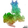
| 手法 | 電子顕微鏡法 / 単粒子再構成法 / クライオ電子顕微鏡法 / 解像度: 3.96 Å | ||||||
 データ登録者 データ登録者 | Yan, Y. / Hardwick, S. / Ron, D. | ||||||
| 資金援助 |  英国, 1件 英国, 1件
| ||||||
 引用 引用 | ジャーナル: Nat Struct Mol Biol / 年: 2021 タイトル: Higher-order phosphatase-substrate contacts terminate the integrated stress response. 著者: Yahui Yan / Heather P Harding / David Ron / 要旨: Many regulatory PPP1R subunits join few catalytic PP1c subunits to mediate phosphoserine and phosphothreonine dephosphorylation in metazoans. Regulatory subunits engage the surface of PP1c, locally ...Many regulatory PPP1R subunits join few catalytic PP1c subunits to mediate phosphoserine and phosphothreonine dephosphorylation in metazoans. Regulatory subunits engage the surface of PP1c, locally affecting flexible access of the phosphopeptide to the active site. However, catalytic efficiency of holophosphatases towards their phosphoprotein substrates remains unexplained. Here we present a cryo-EM structure of the tripartite PP1c-PPP1R15A-G-actin holophosphatase that terminates signaling in the mammalian integrated stress response (ISR) in the pre-dephosphorylation complex with its substrate, translation initiation factor 2α (eIF2α). G-actin, whose essential role in eIF2α dephosphorylation is supported crystallographically, biochemically and genetically, aligns the catalytic and regulatory subunits, creating a composite surface that engages the N-terminal domain of eIF2α to position the distant phosphoserine-51 at the active site. Substrate residues that mediate affinity for the holophosphatase also make critical contacts with eIF2α kinases. Thus, a convergent process of higher-order substrate recognition specifies functionally antagonistic phosphorylation and dephosphorylation in the ISR. | ||||||
| 履歴 |
|
- 構造の表示
構造の表示
| ムービー |
ムービービューア |
|---|---|
| 構造ビューア | 分子: MolmilJmol/JSmol |
- ダウンロードとリンク
-ダウンロード
| PDBx/mmCIF形式 | 7nzm.cif.gz | 221.8 KB | 表示 | PDBx/mmCIF形式 |
|---|---|---|---|---|
| PDB形式 | pdb7nzm.ent.gz | 174.6 KB | 表示 | PDB形式 |
| PDBx/mmJSON形式 | 7nzm.json.gz | ツリー表示 | PDBx/mmJSON形式 | |
| その他 |  その他のダウンロード その他のダウンロード |
-検証レポート
| 文書・要旨 | 7nzm_validation.pdf.gz | 1.3 MB | 表示 | wwPDB検証レポート |
|---|---|---|---|---|
| 文書・詳細版 | 7nzm_full_validation.pdf.gz | 1.3 MB | 表示 | |
| XML形式データ | 7nzm_validation.xml.gz | 51.4 KB | 表示 | |
| CIF形式データ | 7nzm_validation.cif.gz | 76.3 KB | 表示 | |
| アーカイブディレクトリ | https://data.pdbj.org/pub/pdb/validation_reports/nz/7nzmftp://data.pdbj.org/pub/pdb/validation_reports/nz/7nzm | HTTPS FTP |
-関連構造データ


-リンク
 PDBj
PDBj


- 集合体
集合体
| 登録構造単位 | 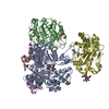
|
|---|---|
| 1 |
|
-要素
-タンパク質 , 5種, 5分子 EBADC
| #1: タンパク質 | 分子量: 21817.863 Da / 分子数: 1 / 変異: phosphorylated Ser51 / 由来タイプ: 組換発現 / 由来: (組換発現) Homo sapiens (ヒト) / 遺伝子: EIF2S1, EIF2A / 発現宿主: |
|---|---|
| #2: タンパク質 | 分子量: 33647.621 Da / 分子数: 1 / 変異: D64A / 由来タイプ: 組換発現 / 由来: (組換発現) 参照: UniProt: P62139, protein-serine/threonine phosphatase |
| #3: タンパク質 | 分子量: 41862.613 Da / 分子数: 1 / 由来タイプ: 天然 / 由来: (天然) |
| #4: タンパク質 | 分子量: 29092.574 Da / 分子数: 1 / 由来タイプ: 天然 / 由来: (天然) |
| #5: タンパク質 | 分子量: 49169.797 Da / 分子数: 1 / 由来タイプ: 組換発現 由来: (組換発現) Homo sapiens (ヒト), (組換発現) 遺伝子: PPP1R15A, GADD34, malE, b4034, JW3994 / 株: K12 / 発現宿主: |
-糖 , 1種, 1分子
| #6: 多糖 | 2-acetamido-2-deoxy-beta-D-glucopyranose-(1-4)-2-acetamido-2-deoxy-beta-D-glucopyranose |
|---|
-非ポリマー , 2種, 2分子 

| #7: 化合物 | ChemComp-MN /  分子量: 54.938 Da / 分子数: 1 / 由来タイプ: 合成 / 式: Mn / タイプ: SUBJECT OF INVESTIGATION 分子量: 54.938 Da / 分子数: 1 / 由来タイプ: 合成 / 式: Mn / タイプ: SUBJECT OF INVESTIGATION |
|---|---|
| #8: 化合物 | ChemComp-ATP /  分子量: 507.181 Da / 分子数: 1 / 由来タイプ: 合成 / 式: C10H16N5O13P3 / コメント: ATP, エネルギー貯蔵分子*YM 分子量: 507.181 Da / 分子数: 1 / 由来タイプ: 合成 / 式: C10H16N5O13P3 / コメント: ATP, エネルギー貯蔵分子*YM |
-詳細
| 研究の焦点であるリガンドがあるか | Y |
|---|
-実験情報
-実験
| 実験 | 手法: 電子顕微鏡法 |
|---|---|
| EM実験 | 試料の集合状態: PARTICLE / 3次元再構成法: 単粒子再構成法 |
- 試料調製
試料調製
| 構成要素 | 名称: pre-dephosphorylation complex of phosphorylated eIF2alpha_2-187 with trapped holophosphatase (PP1A_D64A/PPP1R15A_553-624/G-actin) タイプ: COMPLEX 詳細: One copy of each component was present in the complex: phosphorylated eIF2alpha_2-187, PP1A_D64A, PPP1R15A_553-624, G-actin and DNase I. The full complex was purified by size exclusion chromatography. Entity ID: #1-#5 / 由来: MULTIPLE SOURCES | ||||||||||||||||||||||||||||||||
|---|---|---|---|---|---|---|---|---|---|---|---|---|---|---|---|---|---|---|---|---|---|---|---|---|---|---|---|---|---|---|---|---|---|
| 分子量 | 値: 0.181 MDa / 実験値: YES | ||||||||||||||||||||||||||||||||
| 緩衝液 | pH: 7.4 詳細: 0.22mM Triton X-100 was added into the solution before plunging. | ||||||||||||||||||||||||||||||||
| 緩衝液成分 |
| ||||||||||||||||||||||||||||||||
| 試料 | 濃度: 5 mg/ml / 包埋: NO / シャドウイング: NO / 染色: NO / 凍結: YES | ||||||||||||||||||||||||||||||||
| 試料支持 | 詳細: current 25mA at Pelco EasiGLOW / グリッドの材料: GOLD / グリッドのサイズ: 300 divisions/in. / グリッドのタイプ: UltrAuFoil R0.6/1 | ||||||||||||||||||||||||||||||||
| 急速凍結 | 装置: FEI VITROBOT MARK IV / 凍結剤: ETHANE / 湿度: 100 % / 凍結前の試料温度: 277 K |
- 電子顕微鏡撮影
| 実験機器 |  モデル: Titan Krios / 画像提供: FEI Company |
|---|---|
| 顕微鏡 | モデル: FEI TITAN KRIOS |
| 電子銃 | 電子線源:  FIELD EMISSION GUN / 加速電圧: 300 kV / 照射モード: SPOT SCAN FIELD EMISSION GUN / 加速電圧: 300 kV / 照射モード: SPOT SCAN |
| 電子レンズ | モード: BRIGHT FIELD / 倍率(公称値): 130000 X / 最大 デフォーカス(公称値): -2800 nm / 最小 デフォーカス(公称値): -1000 nm / Cs: 2.7 mm / C2レンズ絞り径: 50 µm |
| 試料ホルダ | 凍結剤: NITROGEN 試料ホルダーモデル: FEI TITAN KRIOS AUTOGRID HOLDER |
| 撮影 | 電子線照射量: 46.84 e/Å2 フィルム・検出器のモデル: GATAN K3 BIOQUANTUM (6k x 4k) 撮影したグリッド数: 1 / 実像数: 4025 |
| 電子光学装置 | エネルギーフィルター名称: GIF Bioquantum / エネルギーフィルタースリット幅: 20 eV / 位相板: VOLTA PHASE PLATE |
- 解析
解析
| EMソフトウェア |
| ||||||||||||||||||||||||||||||||||||
|---|---|---|---|---|---|---|---|---|---|---|---|---|---|---|---|---|---|---|---|---|---|---|---|---|---|---|---|---|---|---|---|---|---|---|---|---|---|
| CTF補正 | 詳細: Warp estimated the CTF parameters and passed them on to CryoSPARC to perform CTF correction. タイプ: PHASE FLIPPING AND AMPLITUDE CORRECTION | ||||||||||||||||||||||||||||||||||||
| 粒子像の選択 | 選択した粒子像数: 132495 | ||||||||||||||||||||||||||||||||||||
| 対称性 | 点対称性: C1 (非対称) | ||||||||||||||||||||||||||||||||||||
| 3次元再構成 | 解像度: 3.96 Å / 解像度の算出法: FSC 0.143 CUT-OFF / 粒子像の数: 60413 / 詳細: non-uniform refinement / 対称性のタイプ: POINT | ||||||||||||||||||||||||||||||||||||
| 原子モデル構築 | B value: 47 / プロトコル: OTHER / 空間: REAL | ||||||||||||||||||||||||||||||||||||
| 原子モデル構築 |
|