 Movie
Movie Controller


+ Open data

- Basic information
| Entry | Database: PDB / ID: 7ngq | |||||||||
|---|---|---|---|---|---|---|---|---|---|---|
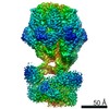
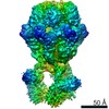
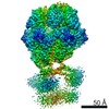
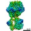
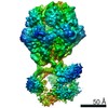
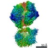
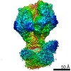
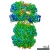
| Title | Human mitochondrial Lon protease homolog, D2-state | |||||||||
 Components Components | Lon protease homolog, mitochondrial | |||||||||
 Keywords Keywords | MOTOR PROTEIN / protease / unfolds / hexamer / AAA+ protein | |||||||||
| Function / homology |  Function and homology information Function and homology informationoxidation-dependent protein catabolic process / PH domain binding / mitochondrial protein catabolic process / G-quadruplex DNA binding / endopeptidase La / mitochondrial DNA metabolic process / mitochondrial genome maintenance / ATP-dependent peptidase activity / protein quality control for misfolded or incompletely synthesized proteins / mitochondrial nucleoid ...oxidation-dependent protein catabolic process / PH domain binding / mitochondrial protein catabolic process / G-quadruplex DNA binding / endopeptidase La / mitochondrial DNA metabolic process / mitochondrial genome maintenance / ATP-dependent peptidase activity / protein quality control for misfolded or incompletely synthesized proteins / mitochondrial nucleoid / insulin receptor substrate binding / chaperone-mediated protein complex assembly / DNA polymerase binding / regulation of peptidyl-tyrosine phosphorylation / negative regulation of insulin receptor signaling pathway / Mitochondrial protein degradation / proteolysis involved in protein catabolic process / mitochondrion organization / ADP binding / protein catabolic process / single-stranded DNA binding / cellular response to oxidative stress / sequence-specific DNA binding / single-stranded RNA binding / response to hypoxia / mitochondrial matrix / serine-type endopeptidase activity / ATP hydrolysis activity / mitochondrion / nucleoplasm / ATP binding / identical protein binding / membrane / cytosol Similarity search - Function | |||||||||
| Biological species |  Homo sapiens (human) Homo sapiens (human) | |||||||||
| Method | ELECTRON MICROSCOPY / single particle reconstruction / cryo EM / Resolution: 12 Å | |||||||||
 Authors Authors | Mohammed, I. / Abrahams, J.P. / Schmitz, K.A. / Maier, T. / Schenck, N. | |||||||||
| Funding support |  Switzerland, 1items Switzerland, 1items
| |||||||||
 Citation Citation | Journal: Structure / Year: 2022 Title: Catalytic cycling of human mitochondrial Lon protease. Authors: Inayathulla Mohammed / Kai A Schmitz / Niko Schenck / Dimitrios Balasopoulos / Annika Topitsch / Timm Maier / Jan Pieter Abrahams / Abstract: The mitochondrial Lon protease (LonP1) regulates mitochondrial health by removing redundant proteins from the mitochondrial matrix. We determined LonP1 in eight nucleotide-dependent conformational ...The mitochondrial Lon protease (LonP1) regulates mitochondrial health by removing redundant proteins from the mitochondrial matrix. We determined LonP1 in eight nucleotide-dependent conformational states by cryoelectron microscopy (cryo-EM). The flexible assembly of N-terminal domains had 3-fold symmetry, and its orientation depended on the conformational state. We show that a conserved structural motif around T803 with a high similarity to the trypsin catalytic triad is essential for proteolysis. We show that LonP1 is not regulated by redox potential, despite the presence of two conserved cysteines at disulfide-bonding distance in its unfoldase core. Our data indicate how sequential ATP hydrolysis controls substrate protein translocation in a 6-fold binding change mechanism. Substrate protein translocation, rather than ATP hydrolysis, is a rate-limiting step, suggesting that LonP1 is a Brownian ratchet with ATP hydrolysis preventing translocation reversal. 3-fold rocking motions of the flexible N-domain assembly may assist thermal unfolding of the substrate protein. | |||||||||
| History |
|
- Structure visualization
| Movie |
Movie viewer |
|---|---|
| Structure viewer | Molecule: MolmilJmol/JSmol |
- Downloads & links
-Download
| PDBx/mmCIF format | 7ngq.cif.gz | 1.5 MB | Display | PDBx/mmCIF format |
|---|---|---|---|---|
| PDB format | pdb7ngq.ent.gz | 1.2 MB | Display | PDB format |
| PDBx/mmJSON format | 7ngq.json.gz | Tree view | PDBx/mmJSON format | |
| Others |  Other downloads Other downloads |
-Validation report
| Summary document | 7ngq_validation.pdf.gz | 1.1 MB | Display | wwPDB validaton report |
|---|---|---|---|---|
| Full document | 7ngq_full_validation.pdf.gz | 1.2 MB | Display | |
| Data in XML | 7ngq_validation.xml.gz | 128.7 KB | Display | |
| Data in CIF | 7ngq_validation.cif.gz | 194.2 KB | Display | |
| Arichive directory | https://data.pdbj.org/pub/pdb/validation_reports/ng/7ngqftp://data.pdbj.org/pub/pdb/validation_reports/ng/7ngq | HTTPS FTP |
-Related structure data
| Related structure data | 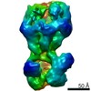 12317MC 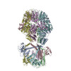 7nfyC 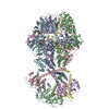 7ng4C 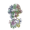 7ng5C 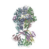 7ngcC 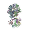 7ngfC 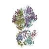 7nglC 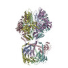 7ngpC  7oxoC M: map data used to model this data C: citing same article ( |
|---|---|
| Similar structure data |


-Links
 PDBj
PDBj
- Assembly
| Deposited unit | 
|
|---|---|
| 1 |
|
-Components
| #1: Protein | Mass: 93201.383 Da / Num. of mol.: 6 Source method: isolated from a genetically manipulated source Source: (gene. exp.) Homo sapiens (human) / Gene: LONP1, PRSS15 / Production host:  #2: Chemical | ChemComp-ADP /   Mass: 427.201 Da / Num. of mol.: 6 / Source method: obtained synthetically / Formula: C10H15N5O10P2 / Feature type: SUBJECT OF INVESTIGATION / Comment: ADP, energy-carrying molecule*YM Mass: 427.201 Da / Num. of mol.: 6 / Source method: obtained synthetically / Formula: C10H15N5O10P2 / Feature type: SUBJECT OF INVESTIGATION / Comment: ADP, energy-carrying molecule*YMHas ligand of interest | Y | |
|---|
-Experimental details
-Experiment
| Experiment | Method: ELECTRON MICROSCOPY |
|---|---|
| EM experiment | Aggregation state: PARTICLE / 3D reconstruction method: single particle reconstruction |
- Sample preparation
| Component | Name: Lon protease homolog, mitochondrial / Type: COMPLEX / Entity ID: #1 / Source: RECOMBINANT |
|---|---|
| Molecular weight | Experimental value: NO |
| Source (natural) | Organism: Homo sapiens (human) |
| Source (recombinant) | Organism: |
| Buffer solution | pH: 7.5 |
| Specimen | Embedding applied: NO / Shadowing applied: NO / Staining applied: NO / Vitrification applied: YES |
| Vitrification | Cryogen name: ETHANE |
- Electron microscopy imaging
| Experimental equipment |  Model: Titan Krios / Image courtesy: FEI Company |
|---|---|
| Microscopy | Model: FEI TITAN KRIOS |
| Electron gun | Electron source:  FIELD EMISSION GUN / Accelerating voltage: 300 kV / Illumination mode: FLOOD BEAM FIELD EMISSION GUN / Accelerating voltage: 300 kV / Illumination mode: FLOOD BEAM |
| Electron lens | Mode: BRIGHT FIELD |
| Image recording | Electron dose: 60 e/Å2 / Detector mode: COUNTING / Film or detector model: GATAN K2 SUMMIT (4k x 4k) |
- Processing
| EM software |
| |||||||||
|---|---|---|---|---|---|---|---|---|---|---|
| CTF correction | Type: PHASE FLIPPING AND AMPLITUDE CORRECTION | |||||||||
| 3D reconstruction | Resolution: 12 Å / Resolution method: FSC 0.143 CUT-OFF / Num. of particles: 11000 / Symmetry type: POINT | |||||||||
| Atomic model building | B value: 850 / Protocol: RIGID BODY FIT / Space: REAL / Target criteria: Correlation coefficient Details: The coordinates of 7ngl subunits were fitted as rigid bodies into the low resolution density of the LonP1 D2-state. The N-domains (res. 123-410) were fitted separately from the combined A- ...Details: The coordinates of 7ngl subunits were fitted as rigid bodies into the low resolution density of the LonP1 D2-state. The N-domains (res. 123-410) were fitted separately from the combined A- and protease domains (res 411-1001). The connecting loops (res 405-415) were reconnected with good geometry. Clashes between the rigid bodies were not corrected due to the low resolution. | |||||||||
| Atomic model building | PDB-ID: 7NGL |
