+ Open data
- Basic information
| Entry | Database: PDB / ID: 7ljr | ||||||
|---|---|---|---|---|---|---|---|
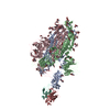
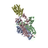
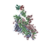
| Title | SARS-CoV-2 Spike Protein Trimer bound to DH1043 fab | ||||||
 Components Components |
| ||||||
 Keywords Keywords | VIRAL PROTEIN/IMMUNE SYSTEM / SARS-CoV-2 Spike Protein Trimer / VIRAL PROTEIN-IMMUNE SYSTEM complex | ||||||
| Function / homology |  Function and homology information Function and homology informationsymbiont-mediated disruption of host tissue / Maturation of spike protein / Translation of Structural Proteins / Virion Assembly and Release / host cell surface / host extracellular space / symbiont-mediated-mediated suppression of host tetherin activity / Induction of Cell-Cell Fusion / structural constituent of virion / membrane fusion ...symbiont-mediated disruption of host tissue / Maturation of spike protein / Translation of Structural Proteins / Virion Assembly and Release / host cell surface / host extracellular space / symbiont-mediated-mediated suppression of host tetherin activity / Induction of Cell-Cell Fusion / structural constituent of virion / membrane fusion / entry receptor-mediated virion attachment to host cell / Attachment and Entry / host cell endoplasmic reticulum-Golgi intermediate compartment membrane / positive regulation of viral entry into host cell / receptor-mediated virion attachment to host cell / host cell surface receptor binding / symbiont-mediated suppression of host innate immune response / endocytosis involved in viral entry into host cell / receptor ligand activity / fusion of virus membrane with host plasma membrane / fusion of virus membrane with host endosome membrane / viral envelope / symbiont entry into host cell / virion attachment to host cell / SARS-CoV-2 activates/modulates innate and adaptive immune responses / host cell plasma membrane / virion membrane / identical protein binding / membrane / plasma membrane Similarity search - Function | ||||||
| Biological species |   Homo sapiens (human) Homo sapiens (human) | ||||||
| Method | ELECTRON MICROSCOPY / single particle reconstruction / cryo EM / Resolution: 3.66 Å | ||||||
 Authors Authors | Gobeil, S. / Acharya, P. | ||||||
| Funding support |  United States, 1items United States, 1items
| ||||||
 Citation Citation |  Journal: bioRxiv / Year: 2021 Journal: bioRxiv / Year: 2021Title: The functions of SARS-CoV-2 neutralizing and infection-enhancing antibodies in vitro and in mice and nonhuman primates. Authors: Dapeng Li / Robert J Edwards / Kartik Manne / David R Martinez / Alexandra Schäfer / S Munir Alam / Kevin Wiehe / Xiaozhi Lu / Robert Parks / Laura L Sutherland / Thomas H Oguin / Charlene ...Authors: Dapeng Li / Robert J Edwards / Kartik Manne / David R Martinez / Alexandra Schäfer / S Munir Alam / Kevin Wiehe / Xiaozhi Lu / Robert Parks / Laura L Sutherland / Thomas H Oguin / Charlene McDanal / Lautaro G Perez / Katayoun Mansouri / Sophie M C Gobeil / Katarzyna Janowska / Victoria Stalls / Megan Kopp / Fangping Cai / Esther Lee / Andrew Foulger / Giovanna E Hernandez / Aja Sanzone / Kedamawit Tilahun / Chuancang Jiang / Longping V Tse / Kevin W Bock / Mahnaz Minai / Bianca M Nagata / Kenneth Cronin / Victoria Gee-Lai / Margaret Deyton / Maggie Barr / Tarra Von Holle / Andrew N Macintyre / Erica Stover / Jared Feldman / Blake M Hauser / Timothy M Caradonna / Trevor D Scobey / Wes Rountree / Yunfei Wang / M Anthony Moody / Derek W Cain / C Todd DeMarco / ThomasN Denny / Christopher W Woods / Elizabeth W Petzold / Aaron G Schmidt / I-Ting Teng / Tongqing Zhou / Peter D Kwong / John R Mascola / Barney S Graham / Ian N Moore / Robert Seder / Hanne Andersen / Mark G Lewis / David C Montefiori / Gregory D Sempowski / Ralph S Baric / Priyamvada Acharya / Barton F Haynes / Kevin O Saunders Abstract: SARS-CoV-2 neutralizing antibodies (NAbs) protect against COVID-19. A concern regarding SARS-CoV-2 antibodies is whether they mediate disease enhancement. Here, we isolated NAbs against the receptor- ...SARS-CoV-2 neutralizing antibodies (NAbs) protect against COVID-19. A concern regarding SARS-CoV-2 antibodies is whether they mediate disease enhancement. Here, we isolated NAbs against the receptor-binding domain (RBD) and the N-terminal domain (NTD) of SARS-CoV-2 spike from individuals with acute or convalescent SARS-CoV-2 or a history of SARS-CoV-1 infection. Cryo-electron microscopy of RBD and NTD antibodies demonstrated function-specific modes of binding. Select RBD NAbs also demonstrated Fc receptor-γ (FcγR)-mediated enhancement of virus infection , while five non-neutralizing NTD antibodies mediated FcγR-independent infection enhancement. However, both types of infection-enhancing antibodies protected from SARS-CoV-2 replication in monkeys and mice. Nonetheless, three of 31 monkeys infused with enhancing antibodies had higher lung inflammation scores compared to controls. One monkey had alveolar edema and elevated bronchoalveolar lavage inflammatory cytokines. Thus, while antibody-enhanced infection does not necessarily herald enhanced infection , increased lung inflammation can occur in SARS-CoV-2 antibody-infused macaques. | ||||||
| History |
|
- Structure visualization
| Movie |
 Movie viewer Movie viewer |
|---|---|
| Structure viewer | Molecule:  Molmil Molmil Jmol/JSmol Jmol/JSmol |
- Downloads & links
- Download
| PDBx/mmCIF format |  7ljr.cif.gz 7ljr.cif.gz | 654.3 KB | Display |  PDBx/mmCIF format PDBx/mmCIF format |
|---|---|---|---|---|
| PDB format |  pdb7ljr.ent.gz pdb7ljr.ent.gz | 515.3 KB | Display |  PDB format PDB format |
| PDBx/mmJSON format |  7ljr.json.gz 7ljr.json.gz | Tree view |  PDBx/mmJSON format PDBx/mmJSON format | |
| Others |  Other downloads Other downloads |
-Validation report
| Arichive directory |  https://data.pdbj.org/pub/pdb/validation_reports/lj/7ljr https://data.pdbj.org/pub/pdb/validation_reports/lj/7ljr ftp://data.pdbj.org/pub/pdb/validation_reports/lj/7ljr ftp://data.pdbj.org/pub/pdb/validation_reports/lj/7ljr | HTTPS FTP |
|---|
-Related structure data
| Related structure data |  23400MC M: map data used to model this data C: citing same article ( |
|---|---|
| Similar structure data |
- Links
Links
- Assembly
| Deposited unit | 
|
|---|---|
| 1 |
|
- Components
| #1: Protein | Mass: 142399.375 Da / Num. of mol.: 3 Source method: isolated from a genetically manipulated source Details: C-terminal tag = T4 fibrin trimerization motif (GYIPEAPRDGQAYVRKDGEWVLLSTFL) + HRV3C protease site (LEVLFQ) + histag (HHHHHHHH) + Strep tag (WSHPQFEKGGGSGGGGSGGSAWSHPQFEK) Source: (gene. exp.)  Gene: S, 2 / Production host:  Homo sapiens (human) / References: UniProt: P0DTC2 Homo sapiens (human) / References: UniProt: P0DTC2#2: Antibody | | Mass: 51632.207 Da / Num. of mol.: 1 Source method: isolated from a genetically manipulated source Source: (gene. exp.)  Homo sapiens (human) / Production host: Homo sapiens (human) / Production host:  Homo sapiens (human) Homo sapiens (human)#3: Antibody | | Mass: 25325.275 Da / Num. of mol.: 1 Source method: isolated from a genetically manipulated source Source: (gene. exp.)  Homo sapiens (human) / Production host: Homo sapiens (human) / Production host:  Homo sapiens (human) Homo sapiens (human) |
|---|
-Experimental details
-Experiment
| Experiment | Method: ELECTRON MICROSCOPY |
|---|---|
| EM experiment | Aggregation state: PARTICLE / 3D reconstruction method: single particle reconstruction |
- Sample preparation
| Component | Name: Spike Protein Trimer bound to the DH1043 antibody / Type: COMPLEX / Entity ID: all / Source: MULTIPLE SOURCES |
|---|---|
| Source (natural) | Organism:  |
| Source (recombinant) | Organism:  Homo sapiens (human) Homo sapiens (human) |
| Details of virus | Empty: NO / Enveloped: YES / Isolate: OTHER / Type: VIRION |
| Buffer solution | pH: 8 |
| Specimen | Embedding applied: NO / Shadowing applied: NO / Staining applied: NO / Vitrification applied: YES |
| Specimen support | Grid type: Quantifoil R1.2/1.3 |
| Vitrification | Cryogen name: ETHANE |
- Electron microscopy imaging
| Experimental equipment |  Model: Titan Krios / Image courtesy: FEI Company |
|---|---|
| Microscopy | Model: FEI TITAN KRIOS |
| Electron gun | Electron source:  FIELD EMISSION GUN / Accelerating voltage: 300 kV / Illumination mode: OTHER FIELD EMISSION GUN / Accelerating voltage: 300 kV / Illumination mode: OTHER |
| Electron lens | Mode: BRIGHT FIELD |
| Image recording | Electron dose: 66.77 e/Å2 / Film or detector model: GATAN K3 (6k x 4k) |
- Processing
| CTF correction | Type: PHASE FLIPPING AND AMPLITUDE CORRECTION |
|---|---|
| 3D reconstruction | Resolution: 3.66 Å / Resolution method: FSC 0.143 CUT-OFF / Num. of particles: 133814 / Symmetry type: POINT |
 Movie
Movie Controller


 PDBj
PDBj


