 Movie
Movie Controller


+ Open data

- Basic information
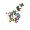
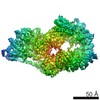
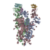
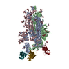
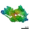
| Entry | Database: PDB / ID: 7l9p | |||||||||||||||||||||
|---|---|---|---|---|---|---|---|---|---|---|---|---|---|---|---|---|---|---|---|---|---|---|
| Title | Structure of human SHLD2-SHLD3-REV7-TRIP13(E253Q) complex | |||||||||||||||||||||
 Components Components |
| |||||||||||||||||||||
 Keywords Keywords | NUCLEAR PROTEIN / REV7 / SHLD2 / SHLD3 / TRIP13 | |||||||||||||||||||||
| Function / homology |  Function and homology information Function and homology informationsomatic diversification of immunoglobulins involved in immune response / DNA damage response, signal transduction resulting in transcription / meiotic recombination checkpoint signaling / negative regulation of ubiquitin protein ligase activity / zeta DNA polymerase complex / synaptonemal complex assembly / positive regulation of isotype switching / : / negative regulation of cell-cell adhesion mediated by cadherin / JUN kinase binding ...somatic diversification of immunoglobulins involved in immune response / DNA damage response, signal transduction resulting in transcription / meiotic recombination checkpoint signaling / negative regulation of ubiquitin protein ligase activity / zeta DNA polymerase complex / synaptonemal complex assembly / positive regulation of isotype switching / : / negative regulation of cell-cell adhesion mediated by cadherin / JUN kinase binding / oocyte maturation / reciprocal meiotic recombination / female meiosis I / negative regulation of epithelial to mesenchymal transition / positive regulation of double-strand break repair via nonhomologous end joining / regulation of double-strand break repair via homologous recombination / mitotic spindle assembly checkpoint signaling / oogenesis / telomere maintenance in response to DNA damage / male meiosis I / positive regulation of peptidyl-serine phosphorylation / spermatid development / error-prone translesion synthesis / negative regulation of double-strand break repair via homologous recombination / site of DNA damage / translesion synthesis / Translesion synthesis by REV1 / Translesion synthesis by POLK / actin filament organization / male germ cell nucleus / Translesion synthesis by POLI / transcription coregulator activity / regulation of cell growth / negative regulation of canonical Wnt signaling pathway / negative regulation of protein catabolic process / spindle / transcription corepressor activity / double-strand break repair / chromosome / actin cytoskeleton / site of double-strand break / spermatogenesis / transcription by RNA polymerase II / RNA polymerase II-specific DNA-binding transcription factor binding / protein-macromolecule adaptor activity / cell division / DNA repair / positive regulation of DNA-templated transcription / chromatin / nucleolus / negative regulation of transcription by RNA polymerase II / ATP hydrolysis activity / nucleoplasm / ATP binding / identical protein binding / nucleus / cytoplasm Similarity search - Function | |||||||||||||||||||||
| Biological species |  Homo sapiens (human) Homo sapiens (human) | |||||||||||||||||||||
| Method | ELECTRON MICROSCOPY / single particle reconstruction / cryo EM / Resolution: 3.6 Å | |||||||||||||||||||||
 Authors Authors | Xie, W. / Patel, D.J. | |||||||||||||||||||||
 Citation Citation | Journal: Proc Natl Acad Sci U S A / Year: 2021 Title: Molecular mechanisms of assembly and TRIP13-mediated remodeling of the human Shieldin complex. Authors: Wei Xie / Shengliu Wang / Juncheng Wang / M Jason de la Cruz / Guotai Xu / Maurizio Scaltriti / Dinshaw J Patel /  Abstract: The Shieldin complex, composed of REV7, SHLD1, SHLD2, and SHLD3, protects DNA double-strand breaks (DSBs) to promote nonhomologous end joining. The AAA ATPase TRIP13 remodels Shieldin to regulate DNA ...The Shieldin complex, composed of REV7, SHLD1, SHLD2, and SHLD3, protects DNA double-strand breaks (DSBs) to promote nonhomologous end joining. The AAA ATPase TRIP13 remodels Shieldin to regulate DNA repair pathway choice. Here we report crystal structures of human SHLD3-REV7 binary and fused SHLD2-SHLD3-REV7 ternary complexes, revealing that assembly of Shieldin requires fused SHLD2-SHLD3 induced conformational heterodimerization of open (O-REV7) and closed (C-REV7) forms of REV7. We also report the cryogenic electron microscopy (cryo-EM) structures of the ATPγS-bound fused SHLD2-SHLD3-REV7-TRIP13 complexes, uncovering the principles underlying the TRIP13-mediated disassembly mechanism of the Shieldin complex. We demonstrate that the N terminus of REV7 inserts into the central channel of TRIP13, setting the stage for pulling the unfolded N-terminal peptide of C-REV7 through the central TRIP13 hexameric channel. The primary interface involves contacts between the safety-belt segment of C-REV7 and a conserved and negatively charged loop of TRIP13. This process is mediated by ATP hydrolysis-triggered rotatory motions of the TRIP13 ATPase, thereby resulting in the disassembly of the Shieldin complex. | |||||||||||||||||||||
| History |
|
- Structure visualization
| Movie |
Movie viewer |
|---|---|
| Structure viewer | Molecule: MolmilJmol/JSmol |
- Downloads & links
-Download
| PDBx/mmCIF format | 7l9p.cif.gz | 545.7 KB | Display | PDBx/mmCIF format |
|---|---|---|---|---|
| PDB format | pdb7l9p.ent.gz | 435.6 KB | Display | PDB format |
| PDBx/mmJSON format | 7l9p.json.gz | Tree view | PDBx/mmJSON format | |
| Others |  Other downloads Other downloads |
-Validation report
| Arichive directory | https://data.pdbj.org/pub/pdb/validation_reports/l9/7l9pftp://data.pdbj.org/pub/pdb/validation_reports/l9/7l9p | HTTPS FTP |
|---|
-Related structure data
| Related structure data |  23244MC  6ww9C  6wwaC M: map data used to model this data C: citing same article ( |
|---|---|
| Similar structure data |


-Links
 PDBj
PDBj


- Assembly
| Deposited unit | 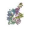
|
|---|---|
| 1 |
|
-Components
| #1: Protein | Mass: 48562.547 Da / Num. of mol.: 6 / Mutation: E253Q Source method: isolated from a genetically manipulated source Source: (gene. exp.) Homo sapiens (human) / Gene: TRIP13, PCH2 / Production host:  #2: Protein | Mass: 24323.348 Da / Num. of mol.: 4 Source method: isolated from a genetically manipulated source Details: closed form / Source: (gene. exp.) Homo sapiens (human) / Gene: MAD2L2, MAD2B, REV7 / Production host: #3: Protein | Mass: 10678.139 Da / Num. of mol.: 2 Fragment: SHLD2 (UNP residues 5-19) + linker + SHLD3 (UNP residues 2-58) Source method: isolated from a genetically manipulated source Source: (gene. exp.) Homo sapiens (human) / Gene: SHLD2, FAM35A, RINN2, SHLD3, FLJ26957, RINN1 / Production host: #4: Chemical | ChemComp-AGS /   Mass: 523.247 Da / Num. of mol.: 5 / Source method: obtained synthetically / Formula: C10H16N5O12P3S / Feature type: SUBJECT OF INVESTIGATION / Comment: ATP-gamma-S, energy-carrying molecule analogue*YM Mass: 523.247 Da / Num. of mol.: 5 / Source method: obtained synthetically / Formula: C10H16N5O12P3S / Feature type: SUBJECT OF INVESTIGATION / Comment: ATP-gamma-S, energy-carrying molecule analogue*YMHas ligand of interest | Y | Has protein modification | N | |
|---|
-Experimental details
-Experiment
| Experiment | Method: ELECTRON MICROSCOPY |
|---|---|
| EM experiment | Aggregation state: PARTICLE / 3D reconstruction method: single particle reconstruction |
- Sample preparation
| Component | Name: SHLD2.3-REV7(4)-TRIP13(E253Q) complex with ATP-gamma-S Type: COMPLEX / Entity ID: #1-#3 / Source: RECOMBINANT |
|---|---|
| Source (natural) | Organism: Homo sapiens (human) |
| Source (recombinant) | Organism: |
| Buffer solution | pH: 7.3 Details: 20 mM HEPES, pH 7.3, 300 mM NaCl, 5 mM MgCl2, 0.1 mM ATP-gamma-S, 1 mM DTT |
| Specimen | Conc.: 0.3 mg/ml / Embedding applied: NO / Shadowing applied: NO / Staining applied: NO / Vitrification applied: YES |
| Specimen support | Grid material: GOLD / Grid type: UltrAuFoil R1.2/1.3 |
| Vitrification | Instrument: FEI VITROBOT MARK IV / Cryogen name: ETHANE / Humidity: 100 % / Chamber temperature: 277 K / Details: 1.5-second blot, blot force of 0 |
- Electron microscopy imaging
| Experimental equipment |  Model: Titan Krios / Image courtesy: FEI Company |
|---|---|
| Microscopy | Model: FEI TITAN KRIOS |
| Electron gun | Electron source:  FIELD EMISSION GUN / Accelerating voltage: 300 kV / Illumination mode: FLOOD BEAM FIELD EMISSION GUN / Accelerating voltage: 300 kV / Illumination mode: FLOOD BEAM |
| Electron lens | Mode: BRIGHT FIELD / Nominal defocus max: -2500 nm / Nominal defocus min: -1000 nm |
| Specimen holder | Cryogen: NITROGEN / Specimen holder model: FEI TITAN KRIOS AUTOGRID HOLDER |
| Image recording | Average exposure time: 0.075 sec. / Electron dose: 53 e/Å2 / Film or detector model: GATAN K3 BIOQUANTUM (6k x 4k) / Num. of grids imaged: 1 / Num. of real images: 40 |
- Processing
| Software | Name: PHENIX / Version: 1.18_3855: / Classification: refinement | ||||||||||||||||||||||||
|---|---|---|---|---|---|---|---|---|---|---|---|---|---|---|---|---|---|---|---|---|---|---|---|---|---|
| EM software | Name: PHENIX / Category: model refinement | ||||||||||||||||||||||||
| CTF correction | Type: PHASE FLIPPING AND AMPLITUDE CORRECTION | ||||||||||||||||||||||||
| Symmetry | Point symmetry: C2 (2 fold cyclic) | ||||||||||||||||||||||||
| 3D reconstruction | Resolution: 3.6 Å / Resolution method: FSC 0.143 CUT-OFF / Num. of particles: 104023 / Symmetry type: POINT | ||||||||||||||||||||||||
| Refine LS restraints |
|
