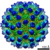
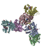
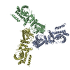
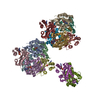
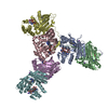
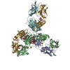
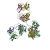
Mouse Norovirus Protruding domain complexed with neutralizing Fab fragment from mAb A6.2
要素
Anti mouse norovirus mAb A6.2 Fab heavy chain
Anti mouse norovirus mAb A6.2 Fab light chain
Capsid protein
キーワード
VIRAL PROTEIN/IMMUNE SYSTEM / Antibody / norovirus / spike / VIRAL PROTEIN / VIRAL PROTEIN-IMMUNE SYSTEM complex
機能・相同性
Calicivirus coat protein C-terminal / Calicivirus coat protein C-terminal / Calicivirus coat protein / Calicivirus coat protein / Picornavirus/Calicivirus coat protein / Viral coat protein subunit / virus-mediated perturbation of host defense response / Capsid protein
National Institutes of Health/National Institute Of Allergy and Infectious Diseases (NIH/NIAID)
1R01-AI141465
米国
引用
ジャーナル: J Virol / 年: 2021 タイトル: A Norovirus Uses Bile Salts To Escape Antibody Recognition While Enhancing Receptor Binding. 著者: Alexis N Williams / Michael B Sherman / Hong Q Smith / Stefan Taube / B Montgomery Pettitt / Christiane E Wobus / Thomas J Smith / 要旨: Noroviruses, members of the family, are the major cause of epidemic gastroenteritis in humans, causing ∼20 million cases annually. These plus-strand RNA viruses have T=3 icosahedral protein ...Noroviruses, members of the family, are the major cause of epidemic gastroenteritis in humans, causing ∼20 million cases annually. These plus-strand RNA viruses have T=3 icosahedral protein capsids with 90 pronounced protruding (P) domain dimers to which antibodies and cellular receptors bind. In the case of mouse norovirus (MNV), bile salts have been shown to enhance receptor (CD300lf) binding to the P domain. We demonstrated previously that the P domains of several genotypes are markedly flexible and "float" over the shell, but the role of this flexibility was unclear. Recently, we demonstrated that bile causes a 90° rotation and collapse of the P domain onto the shell surface. Since bile binds distally to the P-shell interface, it was not at all clear how it could cause such dramatic changes. Here, we present the near-atomic resolution cryo-electron microscopy (cryo-EM) structure of the MNV protruding domain complexed with a neutralizing Fab. On the basis of previous results, we show here that bile salts cause allosteric conformational changes in the P domain that block antibody recognition of the top of the P domain. In addition, bile causes a major rearrangement of the P domain dimers that is likely responsible for the bile-induced collapse of the P domain onto the shell. In the contracted shell conformation, antibodies to the P1 and shell domains are not expected to bind. Therefore, at the site of infection in the gut, the host's own bile allows the virus to escape antibody-mediated neutralization while enhancing cell attachment. The major feature of calicivirus capsids is the 90 protruding domains (P domains) that are the site of cell receptor attachment and antibody epitopes. We demonstrated previously that these P domains are highly mobile and that bile causes these "floating" P domains in mouse norovirus (MNV) to contract onto the shell surface. Here, we present the near-atomic cryo-EM structure of the isolated MNV P domain complexed with a neutralizing Fab fragment. Our data show that bile causes two sets of changes. First, bile causes allosteric conformational changes in the epitopes at the top of the P domain that block antibody binding. Second, bile causes the P domain dimer subunits to rotate relative to each other, causing a contraction of the P domain that buries epitopes at the base of the P and shell domains. Taken together, the results show that MNV uses the host's own metabolites to enhance cell receptor binding while simultaneously blocking antibody recognition.
#256 - 2021年4月 SARSコロナウイルス2型のスパイクと抗体 (SARS-CoV-2 Spike and Antibodies) 類似性 (1)
#156 - 2012年12月 ABO式血液型糖転移酵素 (ABO Blood Type Glycosyltransferases) 類似性 (4)
-
集合体
登録構造単位
A: Capsid protein B: Capsid protein L: Anti mouse norovirus mAb A6.2 Fab light chain W: Anti mouse norovirus mAb A6.2 Fab heavy chain X: Anti mouse norovirus mAb A6.2 Fab light chain Y: Anti mouse norovirus mAb A6.2 Fab heavy chain
 ムービー
ムービー コントローラー


 データを開く
 基本情報
基本情報 要素
要素 キーワード
キーワード 機能・相同性情報
 Murine norovirus 1 (マウスノロウイルス 1)
 データ登録者
 米国, 1件
  引用
引用
 構造の表示
構造の表示 ダウンロードとリンク
 その他のダウンロード


 PDBj
PDBj


 集合体
集合体

 試料調製
試料調製 電子顕微鏡撮影
 FIELD EMISSION GUN / 加速電圧: 300 kV / 照射モード: FLOOD BEAM
 解析
解析