 ムービー
ムービー コントローラー


+ データを開く

- 基本情報
基本情報
| 登録情報 | データベース: PDB / ID: 7kog | |||||||||
|---|---|---|---|---|---|---|---|---|---|---|
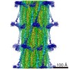
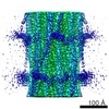
| タイトル | Lethocerus Myosin II complete coiled-coil domain resolved in its native environment | |||||||||
 要素 要素 | Myosin heavy chain isoform Mhc_X1 | |||||||||
 キーワード キーワード | MOTOR PROTEIN / striated muscle / asynchronous flight muscle | |||||||||
| 生物種 |  Lethocerus indicus (昆虫) Lethocerus indicus (昆虫) | |||||||||
| 手法 | 電子顕微鏡法 / 単粒子再構成法 / クライオ電子顕微鏡法 / 解像度: 4.25 Å | |||||||||
 データ登録者 データ登録者 | Rahmani, H. / Hu, Z. / Daneshparvar, N. / Taylor, D. / Taylor, K.A. | |||||||||
| 資金援助 |  米国, 2件 米国, 2件
| |||||||||
 引用 引用 | ジャーナル: Proc Natl Acad Sci U S A / 年: 2021 タイトル: The myosin II coiled-coil domain atomic structure in its native environment. 著者: Hamidreza Rahmani / Wen Ma / Zhongjun Hu / Nadia Daneshparvar / Dianne W Taylor / J Andrew McCammon / Thomas C Irving / Robert J Edwards / Kenneth A Taylor / 要旨: The atomic structure of the complete myosin tail within thick filaments isolated from flight muscle is described and compared to crystal structures of recombinant, human cardiac myosin tail segments. ...The atomic structure of the complete myosin tail within thick filaments isolated from flight muscle is described and compared to crystal structures of recombinant, human cardiac myosin tail segments. Overall, the agreement is good with three exceptions: the proximal S2, in which the filament has heads attached but the crystal structure doesn't, and skip regions 2 and 4. At the head-tail junction, the tail α-helices are asymmetrically structured encompassing well-defined unfolding of 12 residues for one myosin tail, ∼4 residues of the other, and different degrees of α-helix unwinding for both tail α-helices, thereby providing an atomic resolution description of coiled-coil "uncoiling" at the head-tail junction. Asymmetry is observed in the nonhelical C termini; one C-terminal segment is intercalated between ribbons of myosin tails, the other apparently terminating at Skip 4 of another myosin tail. Between skip residues, crystal and filament structures agree well. Skips 1 and 3 also agree well and show the expected α-helix unwinding and coiled-coil untwisting in response to skip residue insertion. Skips 2 and 4 are different. Skip 2 is accommodated in an unusual manner through an increase in α-helix radius and corresponding reduction in rise/residue. Skip 4 remains helical in one chain, with the other chain unfolded, apparently influenced by the acidic myosin C terminus. The atomic model may shed some light on thick filament mechanosensing and is a step in understanding the complex roles that thick filaments of all species undergo during muscle contraction. | |||||||||
| 履歴 |
|
- 構造の表示
構造の表示
| ムービー |
 ムービービューア ムービービューア |
|---|---|
| 構造ビューア | 分子: MolmilJmol/JSmol |
- ダウンロードとリンク
-ダウンロード
| PDBx/mmCIF形式 | 7kog.cif.gz | 414.2 KB | 表示 | PDBx/mmCIF形式 |
|---|---|---|---|---|
| PDB形式 | pdb7kog.ent.gz | 319.3 KB | 表示 | PDB形式 |
| PDBx/mmJSON形式 | 7kog.json.gz | ツリー表示 | PDBx/mmJSON形式 | |
| その他 |  その他のダウンロード その他のダウンロード |
-検証レポート
| 文書・要旨 | 7kog_validation.pdf.gz | 595.6 KB | 表示 | wwPDB検証レポート |
|---|---|---|---|---|
| 文書・詳細版 | 7kog_full_validation.pdf.gz | 628.1 KB | 表示 | |
| XML形式データ | 7kog_validation.xml.gz | 67.1 KB | 表示 | |
| CIF形式データ | 7kog_validation.cif.gz | 102.1 KB | 表示 | |
| アーカイブディレクトリ | https://data.pdbj.org/pub/pdb/validation_reports/ko/7kogftp://data.pdbj.org/pub/pdb/validation_reports/ko/7kog | HTTPS FTP |
-関連構造データ
| 関連構造データ | 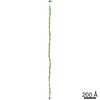 22975MC M: このデータのモデリングに利用したマップデータ C: 同じ文献を引用 ( |
|---|---|
| 電子顕微鏡画像生データ | EMPIAR-10675 (タイトル: The Myosin II Coiled Coil Domain Atomic Structure in its Native Environment Data size: 18.8 TB / Data #1: Frame Stacks 1/3 [micrographs - multiframe] / Data #2: Frame Stacks 2/3 [micrographs - multiframe] / Data #3: Frame Stacks 3/3 [micrographs - multiframe] Data #4: Micrographs before frames alignment [micrographs - single frame] Data #5: Micrographs: frame-aligned and dose-weighted [micrographs - single frame] Data #6: Particle Stack [picked particles - single frame - unprocessed]) |
| 実験データセット #1 | データの種類: EMPIAR / Metadata reference: 10.6019/EMPIAR-10675 |
-リンク
 PDBj
PDBj- 集合体
集合体
| 登録構造単位 | 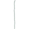
|
|---|---|
| 1 |
|
-要素
| #1: タンパク質 | 分子量: 225227.562 Da / 分子数: 2 / 由来タイプ: 天然 / 由来: (天然) Lethocerus indicus (昆虫) |
|---|
-実験情報
-実験
| 実験 | 手法: 電子顕微鏡法 |
|---|---|
| EM実験 | 試料の集合状態: FILAMENT / 3次元再構成法: 単粒子再構成法 |
- 試料調製
試料調製
| 構成要素 | 名称: Lethocerus flight muscle myosin filament / タイプ: ORGANELLE OR CELLULAR COMPONENT 詳細: The sample is a bipolar helical structure, with helical repeat 145 Angstrom and helical turn 33.98 degree. The sample has C4 symmetry. The map contains 6 unique features: myosin molecule with ...詳細: The sample is a bipolar helical structure, with helical repeat 145 Angstrom and helical turn 33.98 degree. The sample has C4 symmetry. The map contains 6 unique features: myosin molecule with completely resolved rods, 4 resolved non-myosin densities among the myosin rods and an annular region inside of annulus occupied by myosin rods that most likely contains paramyosin. The 4 non-myosin densities may contain parts of the proteins myofilin and flightin. Entity ID: all / 由来: NATURAL |
|---|---|
| 分子量 | 単位: KILODALTONS/NANOMETER / 実験値: NO |
| 由来(天然) | 生物種: Lethocerus indicus (昆虫) / 細胞内の位置: myofibril / 器官: myocyte / Organelle: Sarcomere / 組織: dorsal longitudinal indirect flight muscle |
| 緩衝液 | pH: 6.8 |
| 試料 | 包埋: NO / シャドウイング: NO / 染色: NO / 凍結: YES |
| 急速凍結 | 凍結剤: ETHANE |
- 電子顕微鏡撮影
| 実験機器 |  モデル: Titan Krios / 画像提供: FEI Company |
|---|---|
| 顕微鏡 | モデル: FEI TITAN KRIOS |
| 電子銃 | 電子線源:  FIELD EMISSION GUN / 加速電圧: 300 kV / 照射モード: FLOOD BEAM FIELD EMISSION GUN / 加速電圧: 300 kV / 照射モード: FLOOD BEAM |
| 電子レンズ | モード: BRIGHT FIELD |
| 撮影 | 電子線照射量: 60 e/Å2 / 検出モード: INTEGRATING フィルム・検出器のモデル: DIRECT ELECTRON DE-64 (8k x 8k) 撮影したグリッド数: 1 / 実像数: 3507 |
| 画像スキャン | 横: 8192 / 縦: 8192 / 動画フレーム数/画像: 34 / 利用したフレーム数/画像: 1-34 |
- 解析
解析
| ソフトウェア | 名称: PHENIX / バージョン: 1.17.1_3660: / 分類: 精密化 | ||||||||||||||||||||||||
|---|---|---|---|---|---|---|---|---|---|---|---|---|---|---|---|---|---|---|---|---|---|---|---|---|---|
| EMソフトウェア |
| ||||||||||||||||||||||||
| CTF補正 | タイプ: PHASE FLIPPING AND AMPLITUDE CORRECTION | ||||||||||||||||||||||||
| 3次元再構成 | 解像度: 4.25 Å / 解像度の算出法: FSC 0.143 CUT-OFF / 粒子像の数: 173515 / 対称性のタイプ: POINT | ||||||||||||||||||||||||
| 拘束条件 |
|
