 Movie
Movie Controller

[English] 日本語
 Yorodumi
- PDB-7khw: Cryo-EM structure of enteropathogenic Escherichia coli type III s... -
+ Open data

- Basic information
| Entry | Database: PDB / ID: 7khw | ||||||
|---|---|---|---|---|---|---|---|
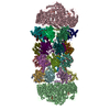
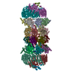
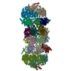
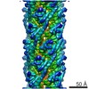
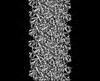
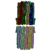
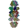
| Title | Cryo-EM structure of enteropathogenic Escherichia coli type III secretion system EspA filament | ||||||
 Components Components | Translocon EspA | ||||||
 Keywords Keywords | PROTEIN FIBRIL / type III secretion system (T3SS) / EspA filament / Helical reconstruction | ||||||
| Function / homology | EspA-like secreted protein / EspA-like secreted protein / EspA/CesA-like / Translocon EspA Function and homology information Function and homology information | ||||||
| Biological species |  | ||||||
| Method | ELECTRON MICROSCOPY / helical reconstruction / cryo EM / Resolution: 3.4 Å | ||||||
 Authors Authors | Zheng, W. / Ilangovan, A. / Costa, T.R.D. / Egelman, E.H. | ||||||
| Funding support |  United States, 1items United States, 1items
| ||||||
 Citation Citation | Journal: Proc Natl Acad Sci U S A / Year: 2021 Title: Cryoelectron-microscopy structure of the enteropathogenic type III secretion system EspA filament. Authors: Weili Zheng / Alejandro Peña / Aravindan Ilangovan / Yasaman Naemi Baghshomali / Gad Frankel / Edward H Egelman / Tiago R D Costa /  Abstract: Enteropathogenic (EPEC) and enterohemorrhagic (EHEC) utilize a macromolecular type III secretion system (T3SS) to inject effector proteins into eukaryotic cells. This apparatus spans the inner and ...Enteropathogenic (EPEC) and enterohemorrhagic (EHEC) utilize a macromolecular type III secretion system (T3SS) to inject effector proteins into eukaryotic cells. This apparatus spans the inner and outer bacterial membranes and includes a helical needle protruding into the extracellular space. Thus far observed only in EPEC and EHEC and not found in other pathogenic Gram-negative bacteria that have a T3SS is an additional helical filament made by the EspA protein that forms a long extension to the needle, mediating both attachment to eukaryotic cells and transport of effector proteins through the intestinal mucus layer. Here, we present the structure of the EspA filament from EPEC at 3.4 Å resolution. The structure reveals that the EspA filament is a right-handed 1-start helical assembly with a conserved lumen architecture with respect to the needle to ensure the seamless transport of unfolded cargos en route to the target cell. This functional conservation is despite the fact that there is little apparent overall conservation at the level of sequence or structure with the needle. We also unveil the molecular details of the immunodominant EspA epitope that can now be exploited for the rational design of epitope display systems. | ||||||
| History |
|
- Structure visualization
| Movie |
Movie viewer |
|---|---|
| Structure viewer | Molecule: MolmilJmol/JSmol |
- Downloads & links
-Download
| PDBx/mmCIF format | 7khw.cif.gz | 1.3 MB | Display | PDBx/mmCIF format |
|---|---|---|---|---|
| PDB format | pdb7khw.ent.gz | 1.1 MB | Display | PDB format |
| PDBx/mmJSON format | 7khw.json.gz | Tree view | PDBx/mmJSON format | |
| Others |  Other downloads Other downloads |
-Validation report
| Arichive directory | https://data.pdbj.org/pub/pdb/validation_reports/kh/7khwftp://data.pdbj.org/pub/pdb/validation_reports/kh/7khw | HTTPS FTP |
|---|
-Related structure data
| Related structure data | 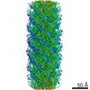 22881MC M: map data used to model this data C: citing same article ( |
|---|---|
| Similar structure data |
-Links
 PDBj
PDBj- Assembly
| Deposited unit | 
|
|---|---|
| 1 |
|
| Symmetry | Helical symmetry: (Circular symmetry: 1 / Dyad axis: no / N subunits divisor: 1 / Num. of operations: 50 / Rise per n subunits: 4.4 Å / Rotation per n subunits: 64.3 °) |
-Components
| #1: Protein | Mass: 20482.811 Da / Num. of mol.: 50 / Source method: isolated from a natural source Source: (natural) Strain: E2348/69 / EPEC / References: UniProt: B7UM94 |
|---|
-Experimental details
-Experiment
| Experiment | Method: ELECTRON MICROSCOPY |
|---|---|
| EM experiment | Aggregation state: FILAMENT / 3D reconstruction method: helical reconstruction |
- Sample preparation
| Component | Name: EspA filament / Type: COMPLEX / Entity ID: all / Source: NATURAL |
|---|---|
| Molecular weight | Experimental value: NO |
| Source (natural) | Organism: Strain: E2348/69 / EPEC |
| Buffer solution | pH: 7.5 |
| Specimen | Embedding applied: NO / Shadowing applied: NO / Staining applied: NO / Vitrification applied: YES |
| Specimen support | Details: unspecified |
| Vitrification | Cryogen name: ETHANE |
- Electron microscopy imaging
| Experimental equipment |  Model: Titan Krios / Image courtesy: FEI Company |
|---|---|
| Microscopy | Model: FEI TITAN KRIOS |
| Electron gun | Electron source:  FIELD EMISSION GUN / Accelerating voltage: 300 kV / Illumination mode: FLOOD BEAM FIELD EMISSION GUN / Accelerating voltage: 300 kV / Illumination mode: FLOOD BEAM |
| Electron lens | Mode: BRIGHT FIELD |
| Specimen holder | Cryogen: NITROGEN |
| Image recording | Electron dose: 55 e/Å2 / Film or detector model: GATAN K3 (6k x 4k) |
- Processing
| EM software |
| ||||||||||||||||||||||||||||||||
|---|---|---|---|---|---|---|---|---|---|---|---|---|---|---|---|---|---|---|---|---|---|---|---|---|---|---|---|---|---|---|---|---|---|
| CTF correction | Type: PHASE FLIPPING AND AMPLITUDE CORRECTION | ||||||||||||||||||||||||||||||||
| Helical symmerty | Angular rotation/subunit: 64.3 ° / Axial rise/subunit: 4.4 Å / Axial symmetry: C1 | ||||||||||||||||||||||||||||||||
| 3D reconstruction | Resolution: 3.4 Å / Resolution method: FSC 0.143 CUT-OFF / Num. of particles: 159460 / Symmetry type: HELICAL |
