 Movie
Movie Controller

[English] 日本語
 Yorodumi
- EMDB-22881: Cryo-EM structure of enteropathogenic Escherichia colitype III se... -
+ Open data

- Basic information
| Entry | Database: EMDB / ID: EMD-22881 | |||||||||
|---|---|---|---|---|---|---|---|---|---|---|
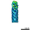
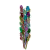
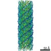
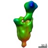
| Title | Cryo-EM structure of enteropathogenic Escherichia colitype III secretion system EspA filament | |||||||||
 Map data Map data | Cryo-EM structure of enteropathogenic Escherichia coli type III secretion system EspA filament | |||||||||
 Sample Sample |
| |||||||||
 Keywords Keywords | type III secretion system (T3SS) / EspA filament / Helical reconstruction / PROTEIN FIBRIL | |||||||||
| Function / homology | EspA-like secreted protein / EspA-like secreted protein / EspA/CesA-like / Translocon EspA Function and homology information Function and homology information | |||||||||
| Biological species |  | |||||||||
| Method | helical reconstruction / cryo EM / Resolution: 3.4 Å | |||||||||
 Authors Authors | Zheng W / Ilangovan A / Costa TRD | |||||||||
| Funding support |  United States, 1 items United States, 1 items
| |||||||||
 Citation Citation | Journal: Proc Natl Acad Sci U S A / Year: 2021 Title: Cryoelectron-microscopy structure of the enteropathogenic type III secretion system EspA filament. Authors: Weili Zheng / Alejandro Peña / Aravindan Ilangovan / Yasaman Naemi Baghshomali / Gad Frankel / Edward H Egelman / Tiago R D Costa /  Abstract: Enteropathogenic (EPEC) and enterohemorrhagic (EHEC) utilize a macromolecular type III secretion system (T3SS) to inject effector proteins into eukaryotic cells. This apparatus spans the inner and ...Enteropathogenic (EPEC) and enterohemorrhagic (EHEC) utilize a macromolecular type III secretion system (T3SS) to inject effector proteins into eukaryotic cells. This apparatus spans the inner and outer bacterial membranes and includes a helical needle protruding into the extracellular space. Thus far observed only in EPEC and EHEC and not found in other pathogenic Gram-negative bacteria that have a T3SS is an additional helical filament made by the EspA protein that forms a long extension to the needle, mediating both attachment to eukaryotic cells and transport of effector proteins through the intestinal mucus layer. Here, we present the structure of the EspA filament from EPEC at 3.4 Å resolution. The structure reveals that the EspA filament is a right-handed 1-start helical assembly with a conserved lumen architecture with respect to the needle to ensure the seamless transport of unfolded cargos en route to the target cell. This functional conservation is despite the fact that there is little apparent overall conservation at the level of sequence or structure with the needle. We also unveil the molecular details of the immunodominant EspA epitope that can now be exploited for the rational design of epitope display systems. | |||||||||
| History |
|
- Structure visualization
| Movie |
Movie viewer |
|---|---|
| Structure viewer | EM map: SurfViewMolmilJmol/JSmol |
| Supplemental images |

- Downloads & links
-EMDB archive
| Map data | emd_22881.map.gz | 59.8 MB | EMDB map data format | |
|---|---|---|---|---|
| Header (meta data) | emd-22881-v30.xmlemd-22881.xml | 10 KB 10 KB | Display Display | EMDB header |
| Images |  emd_22881.png emd_22881.png | 253.4 KB | ||
| Filedesc metadata | emd-22881.cif.gz | 4.9 KB | ||
| Archive directory |  http://ftp.pdbj.org/pub/emdb/structures/EMD-22881ftp://ftp.pdbj.org/pub/emdb/structures/EMD-22881 http://ftp.pdbj.org/pub/emdb/structures/EMD-22881ftp://ftp.pdbj.org/pub/emdb/structures/EMD-22881 | HTTPS FTP |
-Related structure data
| Related structure data | 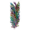 7khwMC M: atomic model generated by this map C: citing same article ( |
|---|---|
| Similar structure data |

-Links
| EMDB pages | EMDB (EBI/PDBe) / EMDataResource |
|---|
-Map
| File | Download / File: emd_22881.map.gz / Format: CCP4 / Size: 64 MB / Type: IMAGE STORED AS FLOATING POINT NUMBER (4 BYTES) | ||||||||||||||||||||||||||||||||||||||||||||||||||||||||||||||||||||
|---|---|---|---|---|---|---|---|---|---|---|---|---|---|---|---|---|---|---|---|---|---|---|---|---|---|---|---|---|---|---|---|---|---|---|---|---|---|---|---|---|---|---|---|---|---|---|---|---|---|---|---|---|---|---|---|---|---|---|---|---|---|---|---|---|---|---|---|---|---|
| Annotation | Cryo-EM structure of enteropathogenic Escherichia coli type III secretion system EspA filament | ||||||||||||||||||||||||||||||||||||||||||||||||||||||||||||||||||||
| Projections & slices | Image control
Images are generated by Spider. | ||||||||||||||||||||||||||||||||||||||||||||||||||||||||||||||||||||
| Voxel size | X=Y=Z: 1.08 Å | ||||||||||||||||||||||||||||||||||||||||||||||||||||||||||||||||||||
| Density |
| ||||||||||||||||||||||||||||||||||||||||||||||||||||||||||||||||||||
| Symmetry | Space group: 1 | ||||||||||||||||||||||||||||||||||||||||||||||||||||||||||||||||||||
| Details | EMDB XML:
CCP4 map header:
| ||||||||||||||||||||||||||||||||||||||||||||||||||||||||||||||||||||
 Z (Sec.)
 Y (Row.)
 X (Col.)


-Supplemental data
- Sample components
-Entire : EspA filament
| Entire | Name: EspA filament |
|---|---|
| Components |
|
-Supramolecule #1: EspA filament
| Supramolecule | Name: EspA filament / type: complex / ID: 1 / Parent: 0 / Macromolecule list: all |
|---|---|
| Source (natural) | Organism: Strain: E2348/69 / EPEC |
-Macromolecule #1: Translocon EspA
| Macromolecule | Name: Translocon EspA / type: protein_or_peptide / ID: 1 / Number of copies: 50 / Enantiomer: LEVO |
|---|---|
| Source (natural) | Organism: Strain: E2348/69 / EPEC |
| Molecular weight | Theoretical: 20.482811 KDa |
| Sequence | String: MDTSTTASVA SANASTSTSM AYDLGSMSKD DVIDLFNKLG VFQAAILMFA YMYQAQSDLS IAKFADMNEA SKESTTAQKM ANLVDAKIA DVQSSSDKNA KAQLPDEVIS YINDPRNDIT ISGIDNINAQ LGAGDLQTVK AAISAKANNL TTTVNNSQLE I QQMSNTLN ...String: MDTSTTASVA SANASTSTSM AYDLGSMSKD DVIDLFNKLG VFQAAILMFA YMYQAQSDLS IAKFADMNEA SKESTTAQKM ANLVDAKIA DVQSSSDKNA KAQLPDEVIS YINDPRNDIT ISGIDNINAQ LGAGDLQTVK AAISAKANNL TTTVNNSQLE I QQMSNTLN LLTSARSDMQ SLQYRTISGI SLGK UniProtKB: Translocon EspA |
-Experimental details
-Structure determination
| Method | cryo EM |
|---|---|
 Processing Processing | helical reconstruction |
| Aggregation state | filament |
-Sample preparation
| Buffer | pH: 7.5 |
|---|---|
| Grid | Pretreatment - Type: PLASMA CLEANING / Details: unspecified |
| Vitrification | Cryogen name: ETHANE |
- Electron microscopy
| Microscope | FEI TITAN KRIOS |
|---|---|
| Image recording | Film or detector model: GATAN K3 (6k x 4k) / Average electron dose: 55.0 e/Å2 |
| Electron beam | Acceleration voltage: 300 kV / Electron source:  FIELD EMISSION GUN FIELD EMISSION GUN |
| Electron optics | Illumination mode: FLOOD BEAM / Imaging mode: BRIGHT FIELD |
| Sample stage | Cooling holder cryogen: NITROGEN |
| Experimental equipment |  Model: Titan Krios / Image courtesy: FEI Company |
-Image processing
| Final reconstruction | Applied symmetry - Helical parameters - Δz: 4.4 Å Applied symmetry - Helical parameters - Δ&Phi: 64.3 ° Applied symmetry - Helical parameters - Axial symmetry: C1 (asymmetric) Resolution.type: BY AUTHOR / Resolution: 3.4 Å / Resolution method: FSC 0.143 CUT-OFF / Software - Name: RELION / Number images used: 159460 |
|---|---|
| Startup model | Type of model: NONE |
| Final angle assignment | Type: NOT APPLICABLE / Software - Name: RELION |
