 ムービー
ムービー コントローラー


+ データを開く

- 基本情報
基本情報
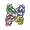
| 登録情報 | データベース: PDB / ID: 7k9l | |||||||||||||||||||||||||||
|---|---|---|---|---|---|---|---|---|---|---|---|---|---|---|---|---|---|---|---|---|---|---|---|---|---|---|---|---|
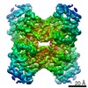
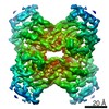
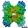
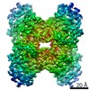
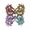
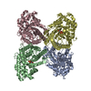
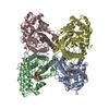
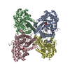
| タイトル | Aldolase, rabbit muscle (no beam-tilt refinement) | |||||||||||||||||||||||||||
 要素 要素 | Fructose-bisphosphate aldolase A | |||||||||||||||||||||||||||
 キーワード キーワード | LYASE / glycolysis / gluconeogenesis / Carbohydrate degradation / Homotetramer | |||||||||||||||||||||||||||
| 機能・相同性 |  機能・相同性情報 機能・相同性情報negative regulation of Arp2/3 complex-mediated actin nucleation / fructose-bisphosphate aldolase / fructose-bisphosphate aldolase activity / M band / I band / glycolytic process / protein homotetramerization / positive regulation of cell migration 類似検索 - 分子機能 | |||||||||||||||||||||||||||
| 生物種 |  | |||||||||||||||||||||||||||
| 手法 | 電子顕微鏡法 / 単粒子再構成法 / クライオ電子顕微鏡法 / 解像度: 4.9 Å | |||||||||||||||||||||||||||
 データ登録者 データ登録者 | Cianfrocco, M.A. / Kearns, S.E. / Cash, J.N. / Li, Y. | |||||||||||||||||||||||||||
| 資金援助 |  米国, 2件 米国, 2件
| |||||||||||||||||||||||||||
 引用 引用 | ジャーナル: IUCrJ / 年: 2020 タイトル: High-resolution cryo-EM using beam-image shift at 200 keV. 著者: Jennifer N Cash / Sarah Kearns / Yilai Li / Michael A Cianfrocco / 要旨: Recent advances in single-particle cryo-electron microscopy (cryo-EM) data collection utilize beam-image shift to improve throughput. Despite implementation on 300 keV cryo-EM instruments, it ...Recent advances in single-particle cryo-electron microscopy (cryo-EM) data collection utilize beam-image shift to improve throughput. Despite implementation on 300 keV cryo-EM instruments, it remains unknown how well beam-image-shift data collection affects data quality on 200 keV instruments and the extent to which aberrations can be computationally corrected. To test this, a cryo-EM data set for aldolase was collected at 200 keV using beam-image shift and analyzed. This analysis shows that the instrument beam tilt and particle motion initially limited the resolution to 4.9 Å. After particle polishing and iterative rounds of aberration correction in , a 2.8 Å resolution structure could be obtained. This analysis demonstrates that software correction of microscope aberrations can provide a significant improvement in resolution at 200 keV. | |||||||||||||||||||||||||||
| 履歴 |
|
- 構造の表示
構造の表示
| ムービー |
ムービービューア |
|---|---|
| 構造ビューア | 分子: MolmilJmol/JSmol |
- ダウンロードとリンク
-ダウンロード
| PDBx/mmCIF形式 | 7k9l.cif.gz | 232.2 KB | 表示 | PDBx/mmCIF形式 |
|---|---|---|---|---|
| PDB形式 | pdb7k9l.ent.gz | 189.7 KB | 表示 | PDB形式 |
| PDBx/mmJSON形式 | 7k9l.json.gz | ツリー表示 | PDBx/mmJSON形式 | |
| その他 |  その他のダウンロード その他のダウンロード |
-検証レポート
| アーカイブディレクトリ | https://data.pdbj.org/pub/pdb/validation_reports/k9/7k9lftp://data.pdbj.org/pub/pdb/validation_reports/k9/7k9l | HTTPS FTP |
|---|
-関連構造データ
| 関連構造データ | 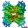 22754MC 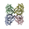 7k9xC  7ka2C 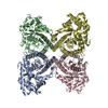 7ka3C 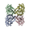 7ka4C M: このデータのモデリングに利用したマップデータ C: 同じ文献を引用 ( |
|---|---|
| 類似構造データ | |
| 電子顕微鏡画像生データ | EMPIAR-10519 (タイトル: Single particle cryo electron microscopy of aldolase (rabbit, muscle) using beam-tilt on Talos Arctica Data size: 87.6 Data #1: Unaligned micrographs of aldolase collected with beam-tilt at 200 kV [micrographs - single frame]) |


-リンク
 PDBj
PDBj
- 集合体
集合体
| 登録構造単位 | 
|
|---|---|
| 1 |
|
-要素
| #1: タンパク質 | 分子量: 39263.672 Da / 分子数: 4 / 由来タイプ: 組換発現 / 由来: (組換発現) Has protein modification | N | |
|---|
-実験情報
-実験
| 実験 | 手法: 電子顕微鏡法 |
|---|---|
| EM実験 | 試料の集合状態: PARTICLE / 3次元再構成法: 単粒子再構成法 |
- 試料調製
試料調製
| 構成要素 | 名称: Homotetramer of aldolase / タイプ: COMPLEX / Entity ID: all / 由来: NATURAL | ||||||||||||
|---|---|---|---|---|---|---|---|---|---|---|---|---|---|
| 分子量 | 値: 157 kDa/nm / 実験値: YES | ||||||||||||
| 由来(天然) | 生物種: | ||||||||||||
| 緩衝液 | pH: 7.5 | ||||||||||||
| 緩衝液成分 |
| ||||||||||||
| 試料 | 濃度: 1.6 mg/ml / 包埋: NO / シャドウイング: NO / 染色: NO / 凍結: YES 詳細: Pure aldolase isolated from rabbit muscle was purchased as a lyophilized powder (Sigma Aldrich) and solubilized in 20 mM HEPES (pH 7.5), 50 mM NaCl at 1.6 mg/ml. Sample was blotted for 4 ...詳細: Pure aldolase isolated from rabbit muscle was purchased as a lyophilized powder (Sigma Aldrich) and solubilized in 20 mM HEPES (pH 7.5), 50 mM NaCl at 1.6 mg/ml. Sample was blotted for 4 seconds with Whatman No. #1 filter paper immediately prior to plunge freezing in liquid ethane cooled by liquid nitrogen. | ||||||||||||
| 試料支持 | グリッドの材料: GOLD / グリッドのサイズ: 300 divisions/in. / グリッドのタイプ: UltrAuFoil R1.2/1.3 | ||||||||||||
| 急速凍結 | 装置: FEI VITROBOT MARK IV / 凍結剤: ETHANE / 湿度: 95 % / 凍結前の試料温度: 277.15 K |
- 電子顕微鏡撮影
| 実験機器 |  モデル: Talos Arctica / 画像提供: FEI Company |
|---|---|
| 顕微鏡 | モデル: FEI TALOS ARCTICA |
| 電子銃 | 電子線源:  FIELD EMISSION GUN / 加速電圧: 200 kV / 照射モード: OTHER FIELD EMISSION GUN / 加速電圧: 200 kV / 照射モード: OTHER |
| 電子レンズ | モード: BRIGHT FIELD / 倍率(公称値): 45000 X / 最大 デフォーカス(公称値): 2 nm / 最小 デフォーカス(公称値): 0.8 nm / Cs: 2.7 mm / C2レンズ絞り径: 100 µm |
| 撮影 | 平均露光時間: 10 sec. / 電子線照射量: 42 e/Å2 フィルム・検出器のモデル: GATAN K2 BASE (4k x 4k) 詳細: Images were collected on a Talos Arctica transmission electron microscope (Thermo Fisher) operating at 200 keV with a gun lens of 6, a spot size of 6, 70 um C2 aperture and 100 um objective ...詳細: Images were collected on a Talos Arctica transmission electron microscope (Thermo Fisher) operating at 200 keV with a gun lens of 6, a spot size of 6, 70 um C2 aperture and 100 um objective aperture using beam-image shift. Movies were collected using a K2 direct electron detector (Gatan Inc.) operating in counting mode at 45,000x corresponding to a physical pixel size of 0.91 A/pixel with a 10 sec exposure using 200 ms per frame. Using an exposure rate of 4.204 e/pix/sec, each movie had a total dose of approximately 42 e/A2 for the 2,111 movies over a defocus 0.8-2 um. |
- 解析
解析
| ソフトウェア | 名称: PHENIX / バージョン: 1.14_3260: / 分類: 精密化 | ||||||||||||||||
|---|---|---|---|---|---|---|---|---|---|---|---|---|---|---|---|---|---|
| EMソフトウェア |
| ||||||||||||||||
| CTF補正 | タイプ: PHASE FLIPPING AND AMPLITUDE CORRECTION | ||||||||||||||||
| 粒子像の選択 | 選択した粒子像数: 718578 | ||||||||||||||||
| 3次元再構成 | 解像度: 4.9 Å / 解像度の算出法: FSC 0.143 CUT-OFF / 粒子像の数: 186471 / 対称性のタイプ: POINT |
